
Presentation
Cerebellar ataxia with spasticity.
Patient Data
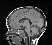

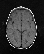
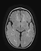
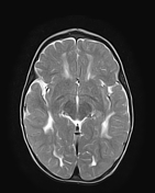
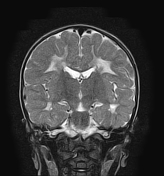
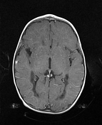
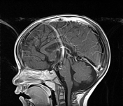
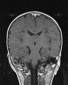
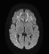
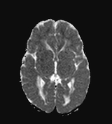

The MRI sequences demonstrate diffuse bilateral, and relatively symmetrical cystic changes of low signal on T1, and FLAIR, and high signal on T2 involving the peri-and juxtaventricular white matter which appears of low signal on T1, and high signal on T2, and FLAIR with no enhancement following IV contrast. The dentate nuclei show also an asymmetrical low signal on T1, high signal FLAIR, and T2 with no enhancement on postcontrast sequences. The corporate-splenial segment of the corpus callosum appears small in size of high signal on FLAIR, and T2 with no enhancement. No involvement of the subcortical arcuate fibres.
Case Discussion
The clinical presentation and the MRI features are suggestive of a vanishing white matter disease, also called childhood ataxia with central hypomyelination (CACH).


 Unable to process the form. Check for errors and try again.