
Presentation
Headache
Patient Data
Age: 35 years
Gender: Female


Show annotations
Download
Info
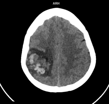
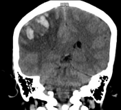
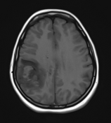
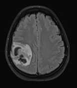
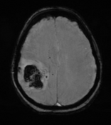
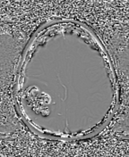
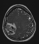
Heterogeneously hyperdense acute haemorrhage in the right high frontoparietal lobes with moderate surrounding oedema.
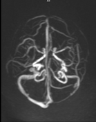
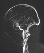
The tubular hyperdense structure in the sulci of the right frontoparietal region could be a cortical vein thrombosis.
Show annotations
Download
Info

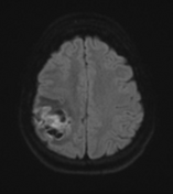
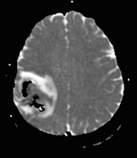
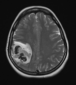
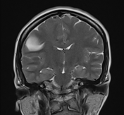
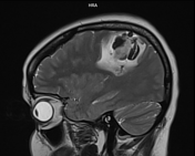
Acute venous haemorrhagic infarct in the right frontoparietal lobes with moderate surrounding oedema. The oedema is larger compared to the size of the haemorrhage, which represents a secondary haemorrhage.
The cortical vein thrombosis in the right frontoparietal region.
Case Discussion
The CT and MRI findings are suggestive venous haemorrhagic infarct due to cortical vein thrombosis.


 Unable to process the form. Check for errors and try again.