
Presentation
Left-sided hemiplegia.
Patient Data
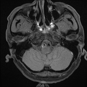

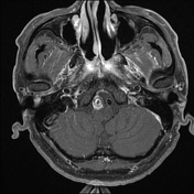
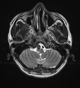
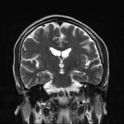
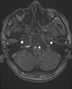
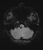

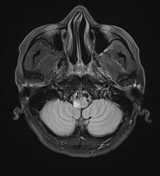
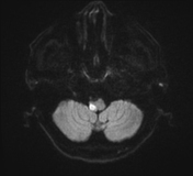
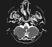
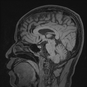

On contrast-enhanced brain MR imaging, an infarct in the right medullary parenchyma was noted, with true diffusion restriction, no haemorrhage, and no post-contrast enhancement.
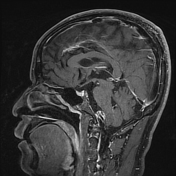
On MRA, moderate to severe stenosis of the proximal V4 segment of the right vertebral artery was observed at the same level as the medullary parenchymal infarct.
On vessel wall MR, there was an dissecting aneurysm of the right vertebral artery and double lumen with intramural haematoma causing corresponding luminal narrowing, scattered high signal on T1W, low signal on SWI, and corresponding high signal on the phase sequence. The intima and outer layers showed strong enhancement post-contrast. Contrast-enhanced T1FS imaging shows strong enhancement of the false lumen, suggesting a T1-contrast effect due to contrast stagnation in the false lumen 1.


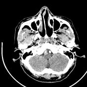
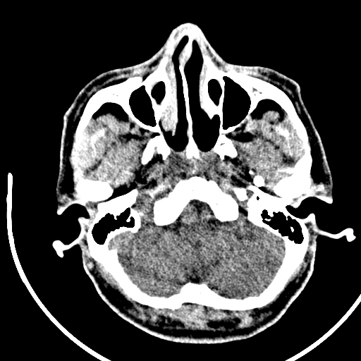
On non-contrast CT, dilation of the proximal V4 segment of the right vertebral artery is observed with increased density. After contrast enhancement, a dissecting aneurysm is noted in the affected vessel segment, with stenosis of the true lumen and strong contrast enhancement in the arterial phase. The false lumen shows marked contrast enhancement in the venous phase, suggesting contrast stagnation, likely due to leakage from the true lumen to the false lumen through an intimal tear.
No abnormalities were noted in the supratentorial and infratentorial brain parenchyma on contrast-enhanced brain CT.
Case Discussion
The findings on brain CT, MRI, and contrast-enhanced vessel wall MRI suggest a vertebral artery dissecting aneurysm with an intramural haematoma causing luminal narrowing, complicated by an infarct at the same level in the medulla.
A study has shown that most patients with vertebral artery dissecting aneurysm (VBAD) demonstrate excellent healing over the follow-up period. The intimal flap in these cases shows wall thickening due to atrophic changes observed on serial HR-VWI (High-Resolution Vessel Wall Imaging) scans. This suggests that conservative management may be effective in treating VBAD in patients with ischaemia and/or headaches. HR-VWI not only benefits the accurate diagnosis of VBAD but also proves useful in monitoring the condition over time, potentially reducing the need for endovascular treatment 1.


 Unable to process the form. Check for errors and try again.