
Presentation
Abdominal mass
Patient Data
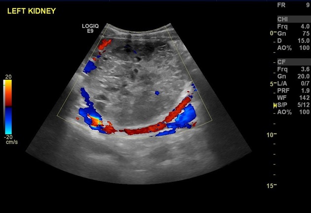
There is a rounded ten by ten centimetres soft tissue mass lesion arising from antero-lateral aspect of left kidney causing moderate hydronephrotic changes suggesting Wilm’s tumour. No lymph node enlargement. CT scan is suggested.
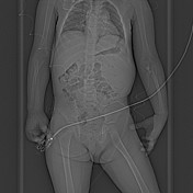
Rest of examination is unremarkable. ( images not included)

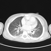

CHEST
Multiple different size small subpleural lung nodules with irregular nodular thickening of the pleura are seen on both sides highly suggestive of lung and pleural metastases.
No evidence of pleural effusion.
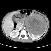
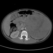
ABDOMEN AND PELVIS
There is a huge heterogeneous, well circumscribed, mass lesion seen arising from the mid and lower poles of the left kidney measuring about twelve centimetres in diameter, crossing the midline displacing the pancreas and bowel loops anteromedially and the aorta to the right side. It appears abutting the psoas muscle
No evidence that the previously described mass is seen invading the veins or evidence of renal vein thrombosis.
This mass lesion is typical for Wilms tumour.
There are multiple enlarged retroperitoneal and mesenteric lymph nodes noted. However no evidence of liver or splenic metastases.
The right kidney and pelvic organs appear normal.
On bone window, there is no evidence of lytic or blastic lesions.
Case Discussion
This is a 5 year old girl, presenting to her primary physician with abdominal mass. Initially was evaluated by ultrasound and the mass was seen arising from the left kidney. Further characterisation and evaluation of this lesion were done by CAT scan with intravenous contrast enhancement. The left renal mass was clearly seen showing typical features of Wilms tumour depicting claw sign and lack of calcification. Unfortunately, lung metastases were identified with enlarged retroperitoneal and mesenteric lymph nodes, but no evidence of bone metastases. It was identified as stage IV disease, unfortunately.


 Unable to process the form. Check for errors and try again.