
Presentation
Patient with a past medical history of IVC and bilateral renal vein thrombosis presents to the ED for gross hematuria.
Patient Data


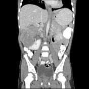
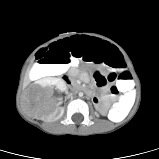
There is a large solid and mildly heterogeneously enhancing mass in the interpolar region of the right kidney with a predominantly expansile growth pattern appearance, though with infiltration of the renal pelvis and probable extension into the right renal vein. The left kidney is atrophic and contains two additional small enhancing and expansile masses.
Note the chronic thrombosis of the IVC and collateral vein formation in the abdomen.
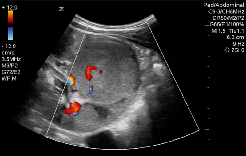
Grey-scale images during an US-guided biopsy demonstrate a large rounded and expansive mass in the right kidney with minimally heterogenous echotexture which is otherwise similar to adjacent renal parenchyma.
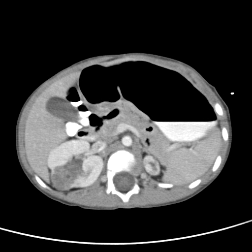
Interval marked decreased size of the bilateral renal masses.
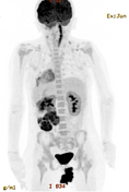
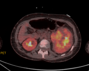
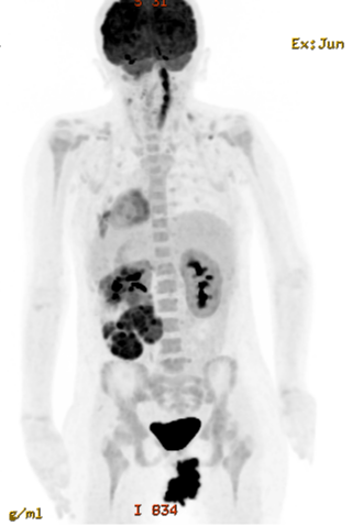
Supplemental case of a 10 year old female.
PET/CT images demonstrate enlargement of the left kidney related to a large, predominantly solid, expansile, and hypermetabolic mass along the inferior renal pole. The mass demonstrates central regions of non-FDG avidity.
Case Discussion
CT performed in the ED demonstrates a large solid and enhancing mass in the interpolar region of the right kidney with a predominantly expansile growth pattern appearance, though with infiltration of the renal pelvis and probable extension into the right renal vein. The left kidney was noted to be atrophic, likely related to his prior history of renal vein thrombosis, but there appear to be 2 additional small enhancing masses in the left kidney, also with an expansive pattern of growth.
Patient subsequently underwent US-guided biopsy which demonstrates a large rounded/expansive mass in the right kidney with minimally heterogenous echotexture which is otherwise similar to adjacent renal parenchyma.
Patient underwent 6 week of chemotherapy. Repeat CECT 2 months later demonstrated marked decreased size of the bilateral renal masses, more notably on the right.
Patient underwent partial right and radical left nephrectomy which confirmed bilateral Wilms tumors.
The supplemental case is of a 10 year old female with a history of biopsy proven Wilms tumor. PET/CT images demonstrate enlargement of the left kidney related to a large, predominantly solid, expansile, and hypermetabolic mass along the inferior renal pole. Central regions of non-FDG avidity suggestive of necrosis were noted.
Key points:
most common pediatric renal mass.
they typically present as an expansile soft tissue mass with patchy enhancement and occasional areas of calcification and macroscopic fat on CT
remember to look for tumor thrombus in the renal vein, IVC, and right atrium
they typically demonstrate hypointense signal on T1-WI, hyperintense signal on T2-WI, and heterogeneous enhancement on MRI


 Unable to process the form. Check for errors and try again.