
Presentation
Distended abdomen in a toddler. Parental concern. Prior ultrasound performed.
Patient Data
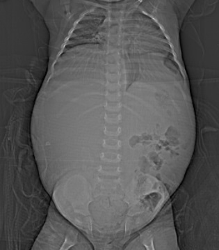
The scanogram from the CT (equating to a plain film) showing a huge homogeneous right sided abdominal mass, crossing the midline and displacing the bowel inferolaterally.
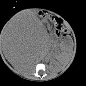


Huge right sided renal derived mass, without internal calcification.
Huge displacement of liver and adjacent structures from right renal mass.
No calcification in the right sided abdominal mass. An important consideration with paediatric abdominal mass in distinguishing a Wilm's tumour from neuroblastoma.
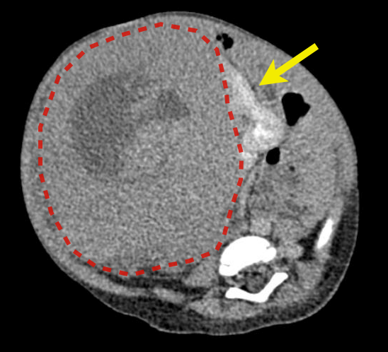
Large mass (Wilm's tumour) outlined by red line with claw sign seen in association with a displaced right kidney (yellow arrow) confirming the renal origin of the mass.
Case Discussion
No calcification in the right sided abdominal mass. An important consideration with paediatric abdominal mass in distinguishing a Wilm's tumour from neuroblastoma, the chief differential in a child of this age.


 Unable to process the form. Check for errors and try again.