
Presentation
Presented for evaluation of fetal congenital anomalies
Patient Data

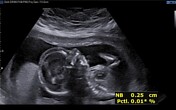
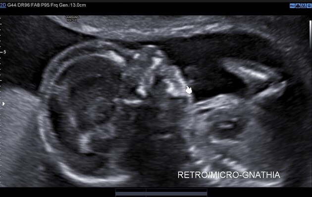
parasagittal view of the fetal face shows evidence of retrognathia/micrognathia
parasagittal view of the fetal face shows evidence of a relatively hypoplastic nasal bone
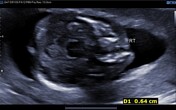
axial view of fetal skull base at the level of hard palate shows evidence of unilateral right cleft lip and palate
coronal view of fetal face shows evidence of unilateral right cleft lip
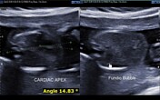
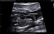
axial views of fetal abdomen and chest shows normal left sided fundic bubble and cardiac apex/axis directed towards left. However, the cardiac axis measured 14.8° suggestive of relative mesocardia
axial view of fetal heart shows abnormal 4 chamber view with hypoplastic left sided cardiac chambers suggestive of hypoplastic left heart syndrome / pulmonary isomerism; right sided cardiac chambers appear normal
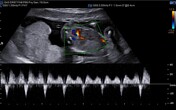
colour doppler flow imaging of the ductus venosus shows evidence of persistent reversal of 'a' wave
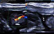
axial view of fetal pelvis shows evidence of single umbilical artery on the right side. Axial section of the umbilical cord shows evidence of absent 'Mickey Mouse sign' suggestive of two vesseled umbilical cord
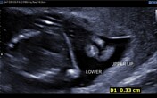
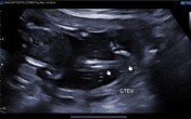
coronal view of fetal leg shows evidence of congenital talipes equinovarus deformity
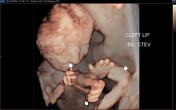
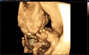
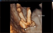
reconstructed 3D image of fetal face and limbs shows evidence of unilateral right cleft lip, retrognathia/ micrognathia and bilateral congenital talipes equinovarus deformities
reconstructed 3D image of the fetal face shows evidence of unilateral right cleft lip
reconstructed 3D image of the fetal legs shows evidence of bilateral congenital talipes equinovarus deformities

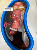
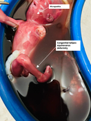
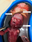
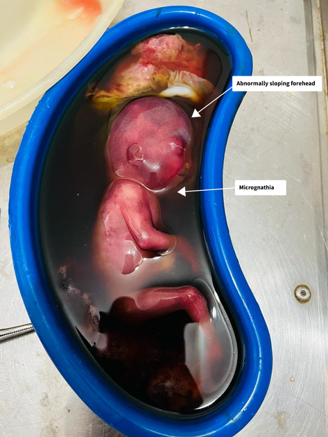
Image (post MTP pathology specimen of the fetus) shows:
evidence of micrognathia/retrognathia and abnormally sloping forehead
an additional finding of poorly formed left external ear pinna with skin flap / tags along its anterior aspect (not detected on ultrasound)
congenital talipes equinovarus deformity
micrognathia/retrognathia, abnormally sloping forehead and unilateral right cleft lip and palate
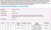
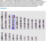
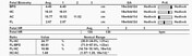
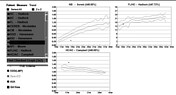

Image:
chromosomal microarray analysis shows a deletion-duplication syndrome, with a 24,291 Kbp (~24.2 MB) deletion on chromosome 4 and 17,237 Kbp (~ 17.2 MB) duplication on chromosome 1, suggesting a chromosomal rearrangement at these loci referred as translocation. CMA analysis shows about 24,291 Kbp (~24.2 MB) deletion on chromosome 4, spanning p16.3-15.2 region. CMA analysis also shows about 17,237 Kbp (~ 17.2 MB) duplication on chromosome 1 spanning q42.2-44, corresponding to partial 1p trisomy syndrome
karyo-view of the above-mentioned findings in image no. 1
detailed fetal biometry shows evidence of asymmetric fetal growth parameters with asymmetric IUGR. Fetal abdominal circumference is significantly reduced with deranged fetal bio-metric ratios i.e. FL/AC, FL/BPD, HC /AC ratios
various fetal bio-metric graphs shows Nasal bone measuring <5th percentile suggestive of hypoplastic nasal bone and asymmetrical IUGR
Case Discussion
A second-time gravida reported for evaluation of fetal congenital anomalies. Gestational age calculated by last menstrual period was 22 weeks and 2 days. The fetus was found to have multiple congenital anomalies during the anomaly scan and was subjected to medical termination of pregnancy. Maternal past history of full-term normal delivery with a healthy baby, now 5 years old. No history of systemic hypertension/pregnancy-induced hypertension or diabetes mellitus type 2 or gestational diabetes mellitus.
Final diagnosis:
A case of Wolf-Hirschhorn syndrome (WHS) with partial trisomy of 1q secondary to deletion-duplication syndrome. A deletion-duplication syndrome, with a ~24.2 MB deletion on chromosome 4 and ~ 17.2 MB duplication on chromosome 1, suggesting translocation.
a. distal deletions of the short arm of chromosome 4, including 4p16.3 micro-deletion syndrome, causes Wolf-Hirschhorn syndrome. The size of the deletion varies among affected individuals; studies suggest that larger deletions tend to result in more severe intellectual disability and physical abnormalities than smaller deletions 1,2.
b. partial duplication on the distal region of the long arm of chromosome 1 is a very rare condition referred to as either pure trisomy or unbalanced translocations, affecting only 200 or so patients worldwide 3.
The fetus in this case had many features of both syndromes.
i. congenital anomalies associated with WHS, that were noted in this fetus are: Prenatal growth retardation (IUGR), facial deformities (Greek warrior helmet appearance: high forehead, prominent glabella, hypertelorism, high-arched eyebrows, protruding eyes and micrognathia), cardiac anomalies- pulmonary isomerism, abnormality of the external ear 1,2.
ii. congenital anomalies associated with partial trisomy of chromosome 1, that were noted in this fetus are protruding forehead and micrognathia.
The prognosis of Wolf-Hirschhorn syndrome is poor. Frequently, this diagnosis is associated with fetal demise or infant death within the first year of life. Individuals who live past the first year of life, have a slow, but constant physical and mental development. About 1/3 of the patients die within the first two years of life due to a heart defect, aspiration pneumonia, or from a seizure. Only symptomatic treatment is available 1,2.
Differential diagnoses:
Pitt-Rogers-Danks syndrome: has features that overlap with those of Wolf-Hirschhorn syndrome. Researchers now recognise that these two conditions are actually part of a single syndrome with variable signs and symptoms
in a fetus with multiple congenital anomalies, other trisomy syndromes such as trisomy 18,13 and 21 also merit consideration
Consent: Before this case was published, the patient and/or their guardian gave written informed consent. Radiopaedia stores this consent separately from this case. If you have any questions, please contact general@radiopaedia.org.
Permission ID: P-00002


 Unable to process the form. Check for errors and try again.