
Yolk sac tumor of infancy
Diagnosis certain
Updates to Case Attributes
Diagnostic Certainty
was set to
.
Race
changed from Caucasian to .
Body
was changed:
SheThe patient underwent surgery but the surgeon wasen seen anydid not see a mass.Eventually vaginoscopy and biopsy was performed performed.
Biopsy proven Yolkyolk sac tumor.
-<p>She underwent surgery but surgeon wasen seen any mass.Eventually vaginoscopy and biopsy was performed.</p><p>Biopsy proven Yolk sac tumor.</p>- +<p>The patient underwent surgery but the surgeon did not see a mass.Eventually vaginoscopy and biopsy was performed.</p><p>Biopsy proven yolk sac tumor.</p>
Systems changed:
- Gynaecology
- Paediatrics
Tags changed:
- germ cell tumour
- uterus
Updates to Study Attributes
Findings
was changed:
In ultrasoundThere is a lesion in the anterior myometriom that is hypervascular and which shows low resistance diastolic flow
Updates to Study Attributes
Findings
was changed:
CT scan shows uterinuterine enlargement which is definitely large for infant,s infants age.
Images Changes:
Image CT (C+ portal venous phase) ( update )
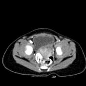
Perspective
was set to
Axial.
Specifics
was set to
C+ portal venous phase.
Image CT (C+ portal venous phase) ( update )
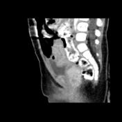
Perspective
was set to
Sagittal.
Specifics
was set to
C+ portal venous phase.
Image CT (C+ portal venous phase) ( update )
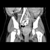
Perspective
was set to
Coronal.
Specifics
was set to
C+ portal venous phase.
Updates to Study Attributes
Findings
was changed:
MRI shows a lobulated mass in the anatomic location of uterus which displays isoisointense signal intensity on the T1 and T2 and slightly hypersignal on the T2 fat sat.
Images Changes:
Image MRI (T1) ( update )
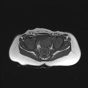
Perspective
was set to
Axial.
Specifics
was set to
T1.
Image MRI (T2 fat sat) ( update )
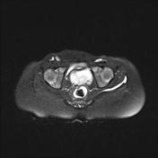
Perspective
was set to
Axial.
Image MRI (T2) ( update )
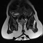
Perspective
was set to
Coronal.
Image MRI (T2) ( update )

Perspective
was set to
Sagittal.
Specifics
was set to
T2.


 Unable to process the form. Check for errors and try again.