
Presentation
Follow-up case of metastatic renal cell carcinoma.
Patient Data
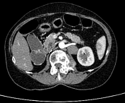

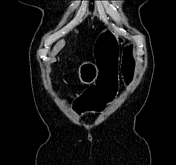
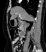
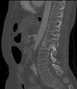
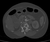
Status post right nephrectomy without any evidence of local recurrence. Two metastatic left paraaortic lymph nodes measuring 23 mm and 11 mm in short axis. Bosniak type 1 left renal measuring 7 mm. Solitary osseous metastasis in the L2 vertebra involving the left half of the body and posterior elements. T10 vertebral body haemangioma.
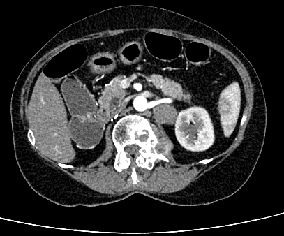
Heterogeneous enhancement pattern of the spleen, which is a normal finding during the arterial phase of the scan.
Case Discussion
During the arterial phase imaging, the spleen shows alternating bands of high and low density resembling the stripes of a zebra, due to its unique vascular anatomy. Assessment of certain splenic pathologies, like splenic laceration or haematoma in the setting of trauma can be challenging if we have only the arterial phase imaging.


 Unable to process the form. Check for errors and try again.