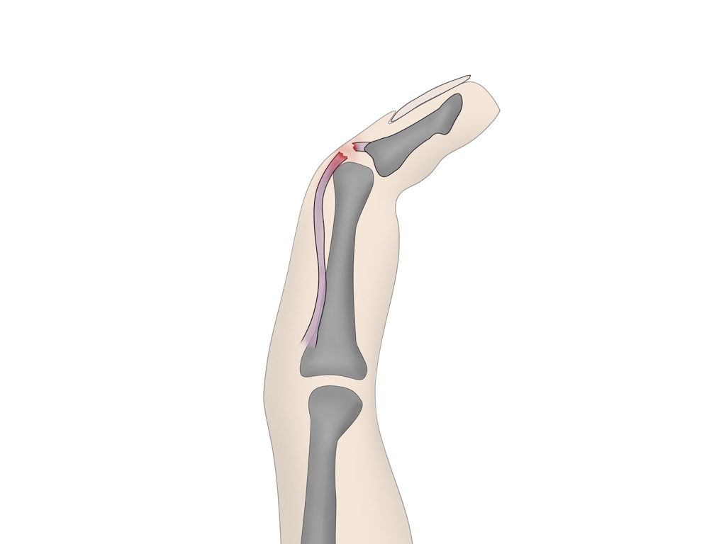
Mallet finger is an injury of the extensor mechanism of the finger at the level of the distal interphalangeal (DIP) joint. It is the most prevalent finger tendon injury in sport. It may represent an isolated tendinous injury or occur in combination with an avulsion fracture of the dorsal base of the distal phalanx.
Clinical presentation
It is characterized by an inability to extend the finger at the distal interphalangeal (DIP) joint. There is slight flexion at this joint, which is where the term "mallet" comes from - the finger position resembles a mallet (for example, a piano key mallet).
The injury classically occurs while playing sports where the DIP joint undergoes sudden flexion (extended finger is struck at the tip by an object, e.g. baseball, basketball), or a crush injury (slamming a door towards the distal interphalangeal joint) in the extensor direction.
Pathology
The DIP joint is extended by combined pulling force of the terminal (lateral bands) of the extensor tendon, functioning together with the oblique retinacular ligament 7. Injury to these structures commonly results from direct axial or flexion loading of the DIP joint, as can occur by direct blow from a ball.
The terminal extensor tendon inserts on the DIP joint capsule, and so injurious force may also result in intra-articular avulsion fracture of the base of the distal phalanx. This may represent an epiphyseal injury in skeletally-immature children 7.
Radiographic features
Plain radiograph
If there is a bony avulsion, a plain film will classically show a triangular avulsion fragment at the insertion of the common extensor tendon on the dorsal aspect of the distal phalanx at the DIP joint.
A high proportion of mallet finger injuries will present as isolated tendon injuries without any associated avulsions fractures known as a "mallet fracture" 5.
Ultrasound
The findings on ultrasound include 6:
loss of real-time movement of the tendon
complete or partial extensor tendon tears
fluid in the region of the extensor tendon insertion
avulsion fracture
Treatment and prognosis
The preferred treatment for closed mallet injuries is non operative treatment, using a splint to maintain the DIP joint in extension or slight hyperextension; the proximal interphalangeal (PIP) joint is kept mobile. This positioning causes approximation of the injured tendon ends, which usually heals by scarring over time and restores extension 7. Non-operative treatment would usually involve 6 weeks of full time splinting followed by 6 weeks of night splinting 8. It is also important to note that the DIP joint should be kept in full extension for the entire first 6 week period including times of hygiene.
It is uncommon for closed mallet finger injuries to require surgical intervention 5. Surgery is considered for avulsion fracture where the fragment is larger than 1/3 of the joint surface and there is more than 2 mm displacement, or there is volar subluxation of the distal phalanx which is not reducible in a splint.
The fragment may be pinned with a Kirschner wire (either percutaneously or following open reduction) or indirectly reduced by "door stop" technique with the DIP joint flexed and a stabilizing Kirschner wire placed through the middle phalanx 7. Post-operative complications, e.g. infection or need for further surgery, are common. The alternative is for fixation with a screw or hook plate if the fragment size will accommodate.
Open injuries are generally surgically explored to evaluate for additional tendinous injury.
Complications
the most common complication in mallet finger injuries are dorsal skin complications (including dorsal ulceration, nail deformities, and maceration of the skin), which are prevalent in both operative and non-operative cases 8
extensor lag
untreated mallet finger or incomplete healing may progress to a swan neck deformity that will require surgical intervention 3,4,7
Avulsion of the flexor digitorum profundus (Jersey finger)
Illustration credit: Andrew Murphy
Jersey finger (also called rugby finger or sweater finger) describes a type of injury where there is avulsion of the flexor digitorum profundus (FDP) from the volar aspect of the distal phalanx base 1. It classically occurs during certain sports resulting from sudden hyperextension of an actively flexed finger (e.g. grabbing an opponent's jersey during rugby or American football.)
It most commonly affects the 4th digit because the FDP insertion into the ring finger is anatomically weaker than the middle finger 2.
Clinical presentation
It is characterized by an inability to flex the finger at the distal interphalangeal (DIP) joint. There is a slight extension at this joint. There is pain and tenderness over the volar distal finger 1.
Radiographs can often be normal 3. If there is a bony avulsion, a triangular avulsion fragment at the volar aspect of the distal phalanx base and overlying soft tissue swelling may be seen.
MRI
Disruption of flexor digitorum profundus (FDP) at the volar base of distal phalanx ± avulsion fragment. MRI also allows visualization of the location of the end of tendons which will affect the surgical classification and management of the patient 4,5.
Treatment and prognosis
conservative for partial tear (i.e. splinting, NSAIDs, physical therapy)
surgical intervention: all complete flexor tendon injuries should be surgically repaired or at least referred to an orthopedic hand surgeon; tendon retraction and time from injury are key 1
Findings: Intraarticular fractures involve the volar aspects of the distal phalangeal bases of the third and fourth digits (middle/long and ring fingers). The fragments appear corticated, indicating subacute to chronic age. Both fragments are proximally displaced. The fracture of the third digit involves half of the articular surface for the distal interphalangeal joint.
Distal phalanx physeal fracture with associated nail bed injury (Seymour fracture)
Illustration credit: Andrew Murphy
The Seymour fracture is a clinically important subtype of mallet finger type injury. The Seymour fracture is comprised of a distal phalanx physeal fracture that has an associated nail bed injury commonly with ungual subluxation.
Clinical presentation
The skeletally-immature patient presents with what clinically appears to be a mallet finger deformity with associated soft tissue trauma at the proximal nail fold. The nail plate may demonstrate obvious signs of avulsion or subluxation, lying superficial to the nail fold or in a more occult situation the nail bed injury may be hidden more proximally and deep to the nail fold.
Pathology
The injury pattern can result from a number of mechanisms, the most common of which include crush injuries, sporting injuries (often hyperflexion) and falls. The injury typically occurs when the distal phalanx of a fully extended digit undergoes forceful flexion or the distal phalanx experiences a crush injury for example in a closing door.
Radiographic features
Plain radiograph
In skeletally-immature individuals, a fracture demonstrating a Salter-Harris type I or Salter-Harris type II pattern through the physis of the distal phalanx or a fracture involving the proximal metaphysis 1-2 mm distal to the epiphyseal plate is seen in conjunction with volar angulation of the diaphysis.
Widening of the physis can be appreciated on AP projections while lateral and oblique projections better demonstrate the aberrant volar angulation.
In many cases, disruption of the overlying soft tissue envelope can be appreciated along with some subcutaneous emphysema. In the absence of nail fold and soft tissue envelope changes, clinical correlation should be recommended to exclude a Seymour fracture.
Since a large number of Seymour fractures are the result of crush injuries, care should be taken to assess for retained radiopaque foreign bodies at the site of the fracture.
Serial imaging studies should consider delayed union, malunion and non-union, in addition to assessment for radiographic changes of osteomyelitis. Growth disturbance of the distal phalanx has also been reported in some cases.
Treatment and prognosis
Early review and treatment by a specialist hand (plastic/orthopedic) surgeon are recommended as delayed treatment has been associated with a high risk of infection.
Open injuries are more prone to soft tissue infection and development of osteomyelitis and thus warrant operative intervention.
The importance of the pattern of injury was first highlighted by the Scottish orthopedic surgeon N Seymour in 1966 5. The seminal article titled "Juxta-epiphyseal fracture of the terminal phalanx of the finger" was published in the Journal of Bone and Joint Surgery 5 and the pattern of injury has subsequently carried the eponymous name 'Seymour fracture'. The pattern of injury is less commonly also called a juxta-epiphyseal fracture of the distal phalanx.
Findings: Fracture is seen at the epiphyseal plate of the distal phalanx of the ring finger with dorsal dislocation of the distal metaphysis, representing Salter-Harris fracture type I.
Case credit: Dr Abdelrashed Abdelmoez Elmelegy Seleem rID: 41965
Key Points
these injuries are often easy to appreciate clinically, however radiographic images of high quality are required to appreciate and detect subtle avulsion injuries
Seymour fractures are important to identify due to the associated nail bed injury, risk of infection is high
Distal phalanx fractures are not all crush injuries. Some very specific injury patterns result in unique abnormalities that are clinically important and have a high enough prevalence that they are worthy of their own page in this pathway. I have broken down this point in the pathway into three sections: first, the distal interphalangeal joint extensor mechanism injury aka 'mallet finger', second the avulsion of the flexor digitorum profundus (Jersey finger), and thirdly the distal phalanx physeal fracture with associated nail bed injury (Seymour fracture). What I think is most important about these particular abnormalities is the mechanism, and understanding how that mechanism results in the injury. Rather than simply saying, this is broken, understanding the journey to that injury is a great memory tool. The mallet finger, for example, is an acute injury to the extensor mechanism of the finger at the distal interphalangeal joint. So patients will often present with a distal interphalangeal joint in fixed flexion.
This is often due to stubbing like mechanisms such as trying to catch a ball and suffering direct axial loading to the distal interphalangeal joint, forcing flexion. This forced flexion will see the extensor tendon which inserts at the distal interphalangeal joint to undergo significant forces and can result in an intra-articular fracture at of the base of the distal phalanx on the dorsal side, the result isn’t always a fracture, however, the clinical presentation of a DIP joint in fixed flexion is.
You can contrast the mallet finger injury to the avulsion of the flexor digitorum profundus AKA jersey finger, In which the mechanism is reversed. The name jersey finger or rugby finger depending on your geographic location originates from the finger being caught in an another player’s jersey and “ripped” or “pulled” and is acutely hyperextended resulting in significant force to the Flexor Digital profundas and an avulsion fracture of the base of the volar aspect of the distal phalanx base. These patients will present with an inability to flex finger joints with significant pain over the dorsal aspect of the distal finger.
In skeletally immature patients, you can get mallet type injuries that result in distal phalanx physeal fracture that have an associated nail bed injury, Aka a Seymour fracture. They will suffer the same mechanisms of injury as the mallet finger; however, the treatment is not as simple, the associated nail bad injury complicates the fracture and warrants surgical repair, it is not an injury you want to miss. Although these three injuries have their own names, it's often best to describe the fracture first. That is why I have put the names in the initial introduction of them as secondary rather than primary. Simply declaring, this patient has a Seymour fracture may not convey this patient has an open nail bed injury, and we should never leave these things up to chance.
During 2024 Radiopaedia.org will be organising a number of additional courses both in Australia and around the world. If you would like to be notified of upcoming courses please fill in the form below.
Updating… Please wait.
Unable to process the form. Check for errors and try again.


 Unable to process the form. Check for errors and try again.