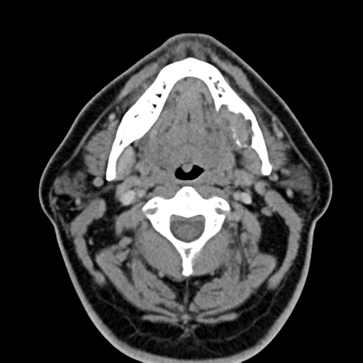
From the case: Ameloblastoma

Axial non-contrast

contributed by Melbourne Uni Radiology Masters on
- “ Pathology MICROSCOPIC DESCRIPTION: Sections show fibroadipose tissue with two lymph nodes. One lymph node contains a 12mm maximum dimension wel...”
View full size version of Ameloblastoma
Updating… Please wait.

 Unable to process the form. Check for errors and try again.
Thank you for updating your details.