From the case: Epidermoid cyst with dystrophic calcification
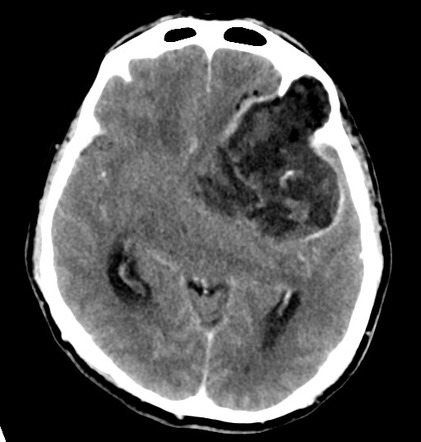
Axial C+ arterial phase

Updating… Please wait.

 Unable to process the form. Check for errors and try again.
Thank you for updating your details.