


1,177 results found
Case
Proximal femur (Gray's illustration)

Published
20 Oct 2020
32% complete
Diagram
Case
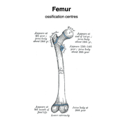
Femur - ossification centers (Gray's illustration)
Published
20 Oct 2020
35% complete
Diagram
Case
Distal femur (Gray's illustration)

Published
20 Oct 2020
32% complete
Diagram
Case
Femur - muscle attachments (Gray's illustration)

Published
20 Oct 2020
32% complete
Diagram
Case
Patella (Gray's illustration)

Published
20 Oct 2020
32% complete
Diagram
Case
Pelvis - muscle attachments (Gray's illustration)

Published
19 Oct 2020
35% complete
Diagram
Case
Hand - ossification centers (Gray's illustrations)

Published
19 Oct 2020
35% complete
Diagram
Case
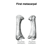
Metacarpals (Gray's illustration)
Published
19 Oct 2020
32% complete
Diagram
Case
Triquetrum (Gray's illustration)

Published
19 Oct 2020
35% complete
Diagram
Case
Trapezium (Gray's illustration)

Published
19 Oct 2020
35% complete
Diagram
Case
Trapezioid (Gray's illustration)

Published
19 Oct 2020
35% complete
Diagram
Case
Scaphoid (Gray's illustration)

Published
19 Oct 2020
35% complete
Diagram
Case
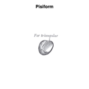
Pisiform (Gray's illustration)
Published
19 Oct 2020
35% complete
Diagram
Case
Lunate (Gray's illustration)

Published
19 Oct 2020
35% complete
Diagram
Case
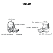
Hamate (Gray's illustration)
Published
19 Oct 2020
35% complete
Diagram
Case
Capitate (Gray's illustration)

Published
19 Oct 2020
35% complete
Diagram
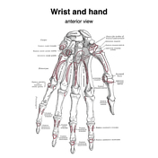
Case
Wrist and hand bones - muscle attachments (Gray's illustration)
Published
16 Oct 2020
35% complete
Diagram
Case
Radius and ulna - muscle attachments (Gray's illustration)

Published
16 Oct 2020
35% complete
Diagram
Case
Ulna - ossification centers (Gray's illustration)

Published
16 Oct 2020
32% complete
Diagram
Case
Radius - ossification centers (Gray's illustration)

Published
16 Oct 2020
32% complete
Diagram
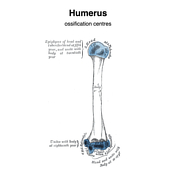
Case
Humerus - ossification centers (Gray's illustrations)
Published
15 Oct 2020
32% complete
Diagram
Case
Scapula - ossification centers (Gray's illustrations)

Published
15 Oct 2020
32% complete
Diagram
Case
Scapula - lateral view (Gray's illustration)

Published
15 Oct 2020
32% complete
Diagram
Case
Humerus - muscle attachments (Gray's illustration)

Published
15 Oct 2020
32% complete
Diagram
Case
Mandibular and dental fracture

Published
13 Oct 2020
85% complete
X-ray
Case
External laryngocoele and pharyngocoele

Published
13 Oct 2020
74% complete
CT
Case
Scapula - muscle attachments (Gray's illustration)

Published
13 Oct 2020
32% complete
Diagram
Case
Mandible - muscle attachments (Gray's illustration)

Published
13 Oct 2020
32% complete
Diagram
Case
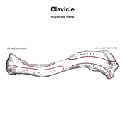
Clavicle - muscle attachments (Gray's illustration)
Published
12 Oct 2020
32% complete
Diagram
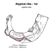
Case
Atypical ribs - 1st and 2nd (Gray's illustration)
Published
12 Oct 2020
35% complete
Diagram
Case
Atypical ribs - 10, 11, 12 (Gray's illustration)

Published
12 Oct 2020
35% complete
Diagram
Case
Typical ribs (Gray's illustration)

Published
12 Oct 2020
35% complete
Diagram
Case
Sternum and costal cartilages - muscle attachments (Gray's illustration)

Published
12 Oct 2020
35% complete
Diagram
Case
Sternum (Gray's illustration)
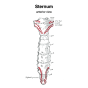
Published
12 Oct 2020
35% complete
Diagram
Case
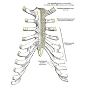
Thoracic cage (Gray's illustrations)
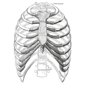
Published
11 Oct 2020
35% complete
Diagram
Case
Lateral spine anatomy (Gray's illustration)
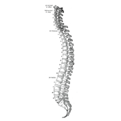
Published
11 Oct 2020
35% complete
Diagram
Case
Vertebral ossification centers
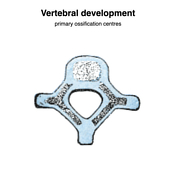
Published
09 Oct 2020
35% complete
Diagram
Case
Sacrum (Gray's illustration)

Published
09 Oct 2020
32% complete
Diagram
Case
Coccyx (Gray's illustration)

Published
09 Oct 2020
32% complete
Diagram
Case
Atypical thoracic vertebrae (Gray's illustration)

Published
08 Oct 2020
35% complete
Diagram
Case
Vertebra prominens (Gray's illustration)

Published
08 Oct 2020
35% complete
Diagram
Case
Atlas (Gray's illustration)

Published
08 Oct 2020
35% complete
Diagram
Case
Axis (Gray's illustration)

Published
08 Oct 2020
35% complete
Diagram
Case
Brainstem arterial territories (diagrams)

Published
07 Oct 2020
44% complete
Diagram
Case
Mandible at different ages (Gray's illustrations)

Published
07 Oct 2020
35% complete
Diagram
Case
Costotransverse joints (Gray's illustration)

Published
07 Oct 2020
35% complete
Diagram
Case
Costovertebral joints (Gray's illustration)

Published
07 Oct 2020
35% complete
Diagram
Case
Costochondral joints (Gray's illustration)
Published
07 Oct 2020
35% complete
Diagram
Case
Typical cervical vertebra (Gray's illustration)

Published
05 Oct 2020
35% complete
Diagram
Case
Typical lumbar vertebra (Gray's illustration)

Published
05 Oct 2020
35% complete
Diagram
Case
Esophageal temperature probe in lung

Published
05 Oct 2020
82% complete
X-ray
Case
Food bolus (lateral neck x-ray)

Published
05 Oct 2020
91% complete
X-ray
Case
Temporomandibular joint anatomy (internal) (Gray's anatomy)

Published
05 Oct 2020
32% complete
Diagram
Case
Temporomandibular joint anatomy (medial) (Gray's anatomy)

Published
05 Oct 2020
35% complete
Diagram
Case
Temporomandibular joint anatomy (lateral) (Gray's anatomy)

Published
05 Oct 2020
32% complete
Diagram
Case
Typical thoracic vertebra (Gray's illustration)

Published
05 Oct 2020
35% complete
Diagram
Case
Accessory articulation of cervical transverse processes

Published
03 Oct 2020
74% complete
Annotated image
CT
Case
Trigeminal nerve cutaneous distribution (Gray's anatomy)

Published
24 Sep 2020
32% complete
Diagram
Case
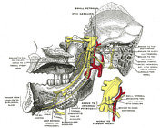
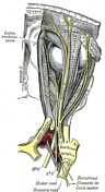
Mandibular division of the trigeminal nerve and submandibular and otic ganglia (Gray's illustration)
Published
20 Sep 2020
35% complete
Diagram
Case
Mandibular division of the trigeminal nerve (Gray's illustration)
Published
20 Sep 2020
35% complete
Diagram
Case
Elbow joint capsule (Gray's illustration)

Published
20 Sep 2020
32% complete
Diagram
Case
Pterygopalatine ganglion

Published
20 Sep 2020
35% complete
Diagram
Case
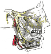
Maxillary division of the trigeminal nerve (Gray's illustration)

Published
20 Sep 2020
35% complete
Diagram
Case
Maxillary and mandibular divisions of the trigeminal nerve (Gray's illustration)
Published
20 Sep 2020
35% complete
Diagram
Case
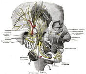
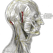
Nerves of the face, scalp and neck (Gray's illustration)
Published
20 Sep 2020
35% complete
Diagram
Case
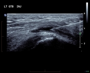
Greater trochanter bursa injection
Published
20 Sep 2020
63% complete
Ultrasound
Case
Anatomy of the genicular ganglion (Gray's illustration)

Published
19 Sep 2020
35% complete
Diagram
Case
Nerves of the orbit (Gray's illustration)

Published
19 Sep 2020
35% complete
Diagram
Case
Anatomy of the ophthalmic division of the trigeminal nerve (Gray's illustration)
Published
19 Sep 2020
35% complete
Diagram
Case
Anatomy of the oculomotor nerve (Gray's illustration)

Published
19 Sep 2020
35% complete
Diagram
Case
Cranial nerve nuclei (axial diagrams)

Published
18 Sep 2020
25% complete
Diagram
Case
Pontine anatomy - CN V (diagram)

Published
18 Sep 2020
32% complete
Diagram
Case
Thrombosed popliteal artery aneurysm and limb ischemia

Published
17 Sep 2020
98% complete
CT
Case
Olfactory nerve (Gray's illustration)

Published
17 Sep 2020
35% complete
Diagram
Case
Optic nerve and chiasm (Gray's illustration)

Published
17 Sep 2020
35% complete
Diagram
Case
Medial elbow ligaments (Gray's illustration)

Published
16 Sep 2020
32% complete
Diagram
Case
Lateral elbow ligaments (Gray's illustration)

Published
16 Sep 2020
35% complete
Diagram
Case
Cranial meninges and falx (Gray's illustration)

Published
14 Sep 2020
35% complete
Diagram
Case
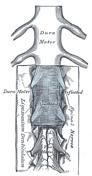
Spinal meninges (Gray's illustration)
Published
14 Sep 2020
35% complete
Diagram
Case
Tentorium cerebelli (Gray's illustration)
Published
14 Sep 2020
32% complete
Diagram
Case
Insular cortex (Gray's illustration)

Published
14 Sep 2020
32% complete
Diagram
Case
Basal ganglia (Gray's illustration)
Published
14 Sep 2020
35% complete
Diagram
Case
Heel soft tissue infection with gas forming organism

Published
13 Sep 2020
88% complete
X-ray
Case
Choledocholithiasis in a patient with left isomerism (polysplenism)

Published
13 Sep 2020
98% complete
X-ray
CT
Case
Brainstem cross-sectional anatomy (diagrams)

Published
08 Sep 2020
29% complete
Diagram
Case
Optic radiations (Gray's illustration)

Published
08 Sep 2020
35% complete
Diagram
Case
Descending spinal tracts (Gray's illustration)

Published
08 Sep 2020
35% complete
Diagram
Case
Ascending spinal tracts (Gray's illustration)

Published
08 Sep 2020
35% complete
Diagram
Case
Cranial nerves (Gray's illustration)

Published
08 Sep 2020
32% complete
Diagram
Case
Spinal cord (Gray's illustration)

Published
07 Sep 2020
35% complete
Diagram
Case
Cauda equina (Gray's illustration)

Published
07 Sep 2020
35% complete
Diagram
Case
Pharyngeal constrictors (Gray's illustration)

Published
07 Sep 2020
35% complete
Diagram
Case
Forearm deep arterial anatomy (Gray's illustration)

Published
06 Sep 2020
35% complete
Diagram
ADVERTISEMENT: Supporters see fewer/no ads