


172 results found
Case
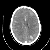
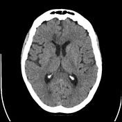
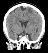
Cortical necrosis post-AMI and cardiac arrest
Published
28 Dec 2023
86% complete
CT
Case
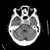
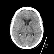
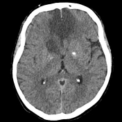
Massive cavernous ICA aneurysm
Published
23 Oct 2023
92% complete
CT
Case
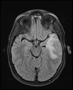
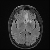
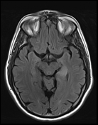
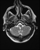
Cerebral abscess secondary to mastoiditis
Published
18 Oct 2023
92% complete
MRI
Case
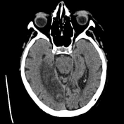
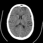
PCA infarction
Published
15 Apr 2023
92% complete
CT
Case
Pericallosal ACA infarction
Published
15 Apr 2023
89% complete
CT
MRI
Case
Gyrus rectus, cingulate gyrus and caudate head infarction post ACA aneurysm coiling
Published
15 Feb 2023
95% complete
MRI
Case
Posterior limb internal capsule infarct after MCA aneurysm coiling
Published
15 Feb 2023
90% complete
CT
Case
Hippocampal and thalamic infarction from vertebral artery dissecting aneursym
Published
15 Feb 2023
84% complete
CT
MRI
Case
Lateral medullary infarct
Published
15 Feb 2023
89% complete
MRI
Case
Bilateral ACA infarction
Published
19 Jan 2023
92% complete
CT
Case
Venous infarct due to superior sagittal sinus and superior cortical vein thrombosis
Published
12 Jan 2023
95% complete
CT
MRI
Case
Bilateral ACA infarction due to azygos ACA embolism

Published
12 Jan 2023
93% complete
MRI
CT
Case
ACA orbitofrontal infarct post DSA
Published
11 Jan 2023
89% complete
CT
DSA (angiography)
Case
Normal head CT venogram
Published
22 Dec 2022
83% complete
CT
Case
Sympathetic nerves (Gray's illustrations)

Published
13 Oct 2022
35% complete
Diagram
Case
Autonomic ganglia of the head and neck (Gray's illustrations)

Published
13 Oct 2022
35% complete
Diagram
Case
Autonomic nervous system (Gray's illustration)

Published
12 Oct 2022
35% complete
Diagram
Case
Bilateral cerebellar tonsil infarction

Published
11 Oct 2022
95% complete
CT
MRI
Case
Tracheal narrowing and deviation from goiter on CXR

Published
04 Oct 2022
82% complete
X-ray
Case
Acute aneurysmal SAH complicated by vasospasm

Published
30 Aug 2022
86% complete
CT
Case
Cervical plexus (Gray's illustrations)

Published
05 Jan 2022
32% complete
Diagram
Case
Brachial plexus (Gray's illustrations)

Published
05 Jan 2022
32% complete
Diagram
Case
Spinal nerve roots (Gray's illustrations)

Published
05 Jan 2022
32% complete
Diagram
Case
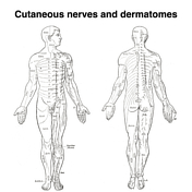
Dermatomes (Gray's illustrations)
Published
05 Jan 2022
35% complete
Diagram
Case
Cutaneous spinal nerves of the upper limb (Gray's illustrations)

Published
05 Jan 2022
29% complete
Diagram
Case
Cutaneous spinal nerves of the lower limb (Gray's illustrations)

Published
05 Jan 2022
29% complete
Diagram
Case
Internal features of the lateral ventricles (Gray's illustrations)

Published
27 Dec 2021
32% complete
Diagram
Case
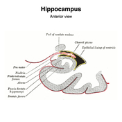
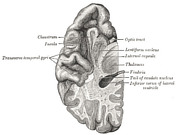
Hippocampus (Gray's illustration)
Published
27 Dec 2021
32% complete
Diagram
Case
Tela choroidea and choroid plexus of lateral ventricles (Gray's illustration)

Published
27 Dec 2021
44% complete
Diagram
Case
Internal capsule fibers (Gray's illustration)

Published
27 Dec 2021
32% complete
Diagram
Case
Fornix (Gray's illustration)

Published
27 Dec 2021
32% complete
Diagram
Case
Corpus striatum (Gray's illustration)

Published
27 Dec 2021
35% complete
Diagram
Case
Corona radiata (Gray's illustration)

Published
27 Dec 2021
32% complete
Diagram
Case
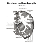
Basal ganglia (Gray's illustrations)
Published
27 Dec 2021
32% complete
Diagram
Case
Dural venous sinuses (Gray's illustrations)

Published
20 Dec 2021
32% complete
Diagram
Case
Cavernous sinus (Gray's illustration)

Published
08 Dec 2021
32% complete
Diagram
Case
Internal cerebral veins (Gray's illustration)

Published
07 Dec 2021
32% complete
Diagram
Case
CT angiogram head sagittal - labeling questions

Published
30 Nov 2021
40% complete
Annotated image
CT
Case
CT angiogram head coronal - labeling questions

Published
29 Nov 2021
40% complete
CT
Annotated image
Case
MRI pituitary gland coronal T1 post contrast - labeling questions

Published
17 Nov 2021
40% complete
Annotated image
MRI
Case
MRI pituitary gland sagittal T1 post contrast - labeling questions

Published
16 Nov 2021
40% complete
MRI
Annotated image
Case
Accessory PCA arising from the terminal ICA

Published
08 Nov 2021
79% complete
MRI
Case
MRI head axial T2 - labeling questions

Published
02 Nov 2021
40% complete
Annotated image
MRI
Case
MRI head sagittal T1 - labeling questions

Published
28 Oct 2021
40% complete
Annotated image
MRI
Case
Superior ophthalmic vein thrombosis

Published
31 Aug 2021
92% complete
CT
Case
Subdural hemorrhage on CT perfusion

Published
26 Aug 2021
39% complete
CT
Case
Toxic encephalopathy

Published
19 May 2021
59% complete
MRI
Case
Toxic encephalopathy

Published
19 May 2021
65% complete
MRI
Case
Subependymal giant cell astrocytoma (SEGA)

Published
03 May 2021
74% complete
MRI
Case
Acquired hepatocerebral degeneration

Published
27 Jan 2021
83% complete
MRI
Case
Cerebral fat embolism

Published
02 Jan 2021
81% complete
CT
X-ray
MRI
Case
CT angiogram head axial - labeling questions

Published
24 Nov 2020
40% complete
CT
Annotated image
Case
Brainstem arterial territories (diagrams)

Published
07 Oct 2020
44% complete
Diagram
Case
Accessory articulation of cervical transverse processes

Published
03 Oct 2020
74% complete
Annotated image
CT
Case
Mandibular division of the trigeminal nerve and submandibular and otic ganglia (Gray's illustration)
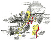
Published
20 Sep 2020
35% complete
Diagram
Case
Mandibular division of the trigeminal nerve (Gray's illustration)
Published
20 Sep 2020
35% complete
Diagram
Case
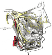
Maxillary division of the trigeminal nerve (Gray's illustration)

Published
20 Sep 2020
35% complete
Diagram
Case
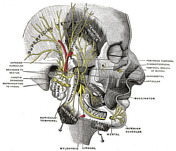
Maxillary and mandibular divisions of the trigeminal nerve (Gray's illustration)
Published
20 Sep 2020
35% complete
Diagram
Case
Nerves of the face, scalp and neck (Gray's illustration)
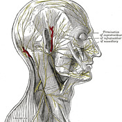
Published
20 Sep 2020
35% complete
Diagram
Case
Anatomy of the genicular ganglion (Gray's illustration)

Published
19 Sep 2020
35% complete
Diagram
Case
Nerves of the orbit (Gray's illustration)

Published
19 Sep 2020
35% complete
Diagram
Case
Anatomy of the ophthalmic division of the trigeminal nerve (Gray's illustration)
Published
19 Sep 2020
35% complete
Diagram
Case
Anatomy of the oculomotor nerve (Gray's illustration)

Published
19 Sep 2020
35% complete
Diagram
Case
Cranial nerve nuclei (axial diagrams)

Published
18 Sep 2020
25% complete
Diagram
Case
Pontine anatomy - CN V (diagram)

Published
18 Sep 2020
32% complete
Diagram
Case
Olfactory nerve (Gray's illustration)

Published
17 Sep 2020
35% complete
Diagram
Case
Optic nerve and chiasm (Gray's illustration)

Published
17 Sep 2020
35% complete
Diagram
Case
Cranial meninges and falx (Gray's illustration)

Published
14 Sep 2020
35% complete
Diagram
Case
Spinal meninges (Gray's illustration)
Published
14 Sep 2020
35% complete
Diagram
Case
Tentorium cerebelli (Gray's illustration)
Published
14 Sep 2020
32% complete
Diagram
Case
Insular cortex (Gray's illustration)

Published
14 Sep 2020
32% complete
Diagram
Case
Basal ganglia (Gray's illustration)
Published
14 Sep 2020
35% complete
Diagram
Case
Brainstem cross-sectional anatomy (diagrams)

Published
08 Sep 2020
29% complete
Diagram
Case
Optic radiations (Gray's illustration)

Published
08 Sep 2020
35% complete
Diagram
Case
Descending spinal tracts (Gray's illustration)

Published
08 Sep 2020
35% complete
Diagram
Case
Ascending spinal tracts (Gray's illustration)

Published
08 Sep 2020
35% complete
Diagram
Case
Cranial nerves (Gray's illustration)

Published
08 Sep 2020
32% complete
Diagram
Case
Spinal cord (Gray's illustration)

Published
07 Sep 2020
35% complete
Diagram
Case
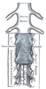
Cauda equina (Gray's illustration)

Published
07 Sep 2020
35% complete
Diagram
Case
Circle of Willis (Gray's illustration)

Published
06 Sep 2020
32% complete
Diagram
Case
Spinal cord cross section (Gray's illustration)

Published
06 Sep 2020
35% complete
Diagram
Case
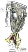
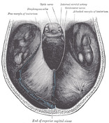
Cranial nerves in the posterior fossa (Gray's illustration)

Published
06 Sep 2020
35% complete
Diagram
Case
Brainstem tracts (Gray's illustrations)

Published
06 Sep 2020
32% complete
Diagram
Case
Pyramids and olives (Gray's illustration)
Published
03 Sep 2020
35% complete
Diagram
Case
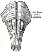
Cerebellar peduncles (Gray's illustration)

Published
03 Sep 2020
35% complete
Diagram
Case
Colliculi connections (Gray's illustration)

Published
03 Sep 2020
35% complete
Diagram
Case
Interconnection of cranial nerve nuclei (Gray's illustration)

Published
03 Sep 2020
35% complete
Diagram
Case
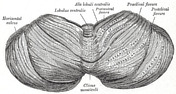
Cerebellum inferior surface (Gray's illustration)

Published
03 Sep 2020
35% complete
Diagram
Case
Cerebellum superior surface (Gray's illustration)
Published
03 Sep 2020
32% complete
Diagram
Case
Cerebellar peduncles (Gray's illustration)

Published
03 Sep 2020
35% complete
Diagram
Case
Dentate nucleus (Gray's illustration)

Published
03 Sep 2020
32% complete
Diagram
Case
Anatomy of the lateral ventricles (Gray's illustration)

Published
03 Sep 2020
32% complete
Diagram
Case
Upper-level pons (Gray's illustration)

Published
03 Sep 2020
32% complete
Diagram
ADVERTISEMENT: Supporters see fewer/no ads


 Unable to process the form. Check for errors and try again.