Anatomy - Neuro, Head & Neck
Playlist contributed by
Yaïr Glick
◉
Play
Share
Playlist
Full screen playlist
Playlist with hidden diagnosis
Full screen playlist with hidden diagnosis
Playlist information
Playlist created:
19 May 2017 by
Yaïr Glick
◉
Last edited:
29 Oct 2023
Number of cases:
49
Number of slides:
0
rID:
135798
Systems:
Central Nervous System
,
Head & Neck
,
Musculoskeletal
,
Spine
,
Vascular
Visibility:
public
Show case titles
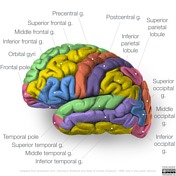
Case 1
Brain lobes - annotated MRI
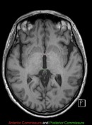
Case 2
Neuroanatomical landmarks
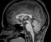
Case 3
Sagittal midline of the brain - normal anatomy
Case 4
CT head sagittal - labeling questions
Case 5
Anatomy: sulci of the brain
Case 6
Neuroanatomy: lateral cortex (diagrams)
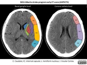
Case 7
MCA - Alberta stroke program early CT score (ASPECTS) illustration
Case 8
Posterior circulation - Acute stroke prognosis early CT score (pc-ASPECTS) illustration
Case 9
Middle cerebral artery territory in ASPECTS study
Case 10
Normal MRA brain (anatomy quiz)
Case 11
Brain venous vascular territories (diagram)
Case 12
Posterior fossa vascular territories (illustration)
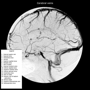
Case 13
Cerebral veins (annotated DSA)
Case 14
Carotid artery segments (diagram)
Case 15
Head and neck vessels (illustrations)
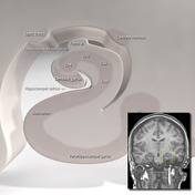
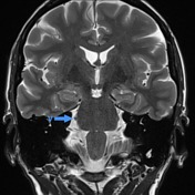
Case 16
Hippocampal anatomy (illustration)
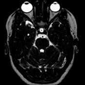
Case 17
Normal MRI epilepsy protocol
Case 18
Normal pituitary MRI (sagittal T2 only)
Case 19
Orbital apex (diagram)
Case 20
Ocular globe (illustration)
Case 21
Normal cranial nerves
Case 22
Trigeminal nerve (normal)
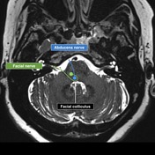
Case 23
Abducens and facial cranial nerves and nuclei
Case 24
Facial nerve anatomy - labeled CT
Case 25
MRI anatomy of the hypoglossal canal and the jugular foramen
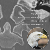
Case 26
Hypoglossal canal (eagle sign)
Case 27
Pterygopalatine fossa (annotated CT)
Case 28
CT facial bones/orbits axial - labeling questions
Case 29
CT facial bones/orbits sagittal - labeling questions
Case 30
Temporal bone divisions: annotated CT
Case 31
Normal petrous temporal bone axial CT - with labels
Case 32
Inner ear anatomy - annotated CT
Case 33
Middle ear anatomy - annotated CT
Case 34
External ear anatomy: annotated CT
Case 35
Brainstem arterial territories (diagrams)
Case 36
Muscles of mastication (annotated image)
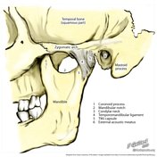
Case 37
Temporomandibular joint (illustration)
Case 38
Temporomandibular joint (illustration)
Case 39
Basilar invagination measurements
Case 40
CT neck with annotated scrollable images
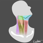
Case 41
Deep spaces of the head and neck - annotated MRI
Case 42
Lymph node levels of the head and neck (annotated CT)
Case 43
Lymph node levels (illustration)
Case 44
Lymph node levels of the neck
Case 45
Vertebral artery
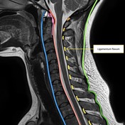
Case 46
Ligaments of the cervical spine (annotated image)
Case 47
Normal ostiomeatal complex (illustration)
Case 48
Park grading system for cervical foraminal stenosis
Case 49
Larynx - coronal CT labeled anatomy