Head and neck
Playlist contributed by
Robert Ligetfalvi
Play
Share
Playlist
Full screen playlist
Playlist with hidden diagnosis
Full screen playlist with hidden diagnosis
Playlist information
Playlist created:
27 Nov 2017 by
Robert Ligetfalvi
Last edited:
27 Nov 2017
Number of cases:
122
Number of slides:
0
rID:
121975
Systems:
Central Nervous System
,
Chest
,
Forensic
,
Gastrointestinal
,
Head & Neck
,
Musculoskeletal
,
Oncology
,
Paediatrics
,
Spine
,
Trauma
,
Vascular
Visibility:
public
Show case titles
Case 1
Pituitary macroadenoma - cavernous sinus invasion
Case 2
Pituitary macroadenoma - necrotic
Case 3
Hemorrhagic pituitary macroadenoma
Case 4
Glioblastoma NOS (extending into ventricle)
Case 5
Subarachnoid hemorrhage
Case 6
Subdural hemorrhage - acute on warfarin
Case 7
Acute infarction
Case 8
Ischemic stroke
Case 9
Hyperdense MCA sign
Case 10
PCA territory infarct
Case 11
Thalamic hemorrhage
Case 12
Diagram - intracranial hemorrhage
Case 13
Extradural hematoma
Case 14
Extradural hematoma, subdural hematomas, cerebral contusions and subgaleal hematoma
Case 15
Extradural hemorrhage
Case 16
Acute left otitis externa - swimmer's ear
Case 17
Necrotizing otitis externa
Case 18
Chronic otitis media
Case 19
Cholesteatoma
Case 20
Acoustic schwannoma (recurrent)
Case 21
Le Fort fracture - type I
Case 22
Chronic maxillary sinusitis
Case 23
Ethmoid mucocoele
Case 24
Carotid body tumor
Case 25
Fibrous dysplasia
Case 26
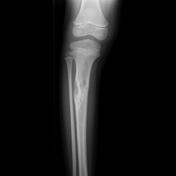
Fibrous dysplasia - tibia
Case 27
Optic neuritis (multiple sclerosis)
Case 28
Retropharyngeal abscess - extensive
Case 29
Supraglottic retropharyngeal abscess
Case 30
Left vocal cord paralysis secondary to pseudocoarctation of the aorta
Case 31
Laryngocele - internal
Case 32
Upper esophageal diverticulum
Case 33
Esophageal adenocarcinoma
Case 34
Esophageal adenocarcinoma (T3N1)
Case 35
Wilson disease
Case 36
Herpes simplex encephalitis
Case 37
Toxic leukoencephalopathy - methotrexate
Case 38
Trans-scaphoid perilunate dislocation
Case 39
Communicating hydrocephalus
Case 40
Brain ventricle anatomy (illustration)
Case 41
Hydrocephalus
Case 42
Pilocytic astrocytoma
Case 43
Optic pathway glioma - NF1
Case 44
Optic nerve glioma
Case 45
Posterior fossa ependymoma
Case 46
Supratentorial ependymoma
Case 47
Medulloblastoma
Case 48
Medulloblastoma - adult
Case 49
Choroid plexus carcinoma
Case 50
Choroid plexus papilloma
Case 51
Hill-Sachs lesion
Case 52
Hill-Sachs lesion
Case 53
Cerebral vein thrombosis
Case 54
Chiari 1 malformation with focal syrinx
Case 55
Chiari 1 malformation with syrinx
Case 56
Klippel-Feil syndrome
Case 57
Acute infarction
Case 58
PCA territory infarct
Case 59
Pontine infarct
Case 60
Fogging phenomenon - stroke
Case 61
Middle cerebral artery territory infarct
Case 62
MRI in hyperacute stroke
Case 63
Subacute cerebral infarction - with MR spectroscopy
Case 64
Alzheimer disease
Case 65
Alzheimer disease
Case 66
Frontal horn, intercaudate and inner table ratios (diagram)
Case 67
Loss of swallow tail sign in Parkinson disease
Case 68
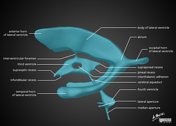
Substantia nigra anatomy
Case 69
Cerebral abscess
Case 70
Cerebral abscess post cardiac surgery
Case 71
Tension pneumocephalus secondary to frontal sinus fracture
Case 72
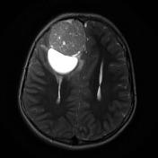
Pilocytic astrocytoma
Case 73
Anaplastic astrocytoma (WHO grade III)
Case 74
Tuberous sclerosis
Case 75
Posterior fossa ependymoma
Case 76
Medulloblastoma - adult
Case 77
Choroid plexus papilloma
Case 78
Concha bullosa - bilateral
Case 79
Frontal mucocele
Case 80
Thyroglossal duct cyst
Case 81
Branchial cleft cyst
Case 82
Carotid body tumor
Case 83
Normal posterior pituitary bright spot
Case 84
Empty sella
Case 85
Empty sella
Case 86
Rathke's cleft cyst
Case 87
Pituitary microadenoma
Case 88
Adamantinomatous craniopharyngioma
Case 89
Necrotizing otitis externa
Case 90
Recurrent cholesteatoma
Case 91
Acoustic schwannoma (large with cystic change)
Case 92
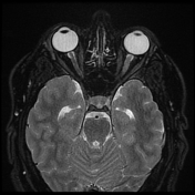
Retinoblastoma
Case 93
Mucocele of the left maxillary antrum
Case 94
Thyroglossal duct cyst
Case 95
Laryngocele - external
Case 96
Distribution of paragangliomas (diagram)
Case 97
Carotid body tumor
Case 98
Carotid body tumor on angiography
Case 99
Normal CT cerebral arteriography
Case 100
Empty sella
Case 101
Haller cell and concha bullosa
Case 102
Chronic maxillary sinusitis
Case 103
Mucoperiosteal hypertrophy: chronic sinusitis
Case 104
Chronic maxillary sinusitis
Case 105
Obstructive hydrocephalus
Case 106
Aqueduct stenosis
Case 107
Acute cerebral infarct with hyperdense MCA sign
Case 108
Base of skull lines (annotated images)
Case 109
Diagram - intracranial hemorrhage
Case 110
Hippocampal sclerosis
Case 111
Mesial temporal sclerosis
Case 112
Temporal lobe atrophy
Case 113
Mesial temporal sclerosis - left
Case 114
Mesial temporal sclerosis
Case 115
Alzheimer disease
Case 116
Ventricles with cava
Case 117
Cavum septum pellucidum
Case 118
Cavum septum pellucidum (gross pathology)
Case 119
Cavum septum pellucidum, cavum vergae, and cavum veli interpositi (annotated CT)
Case 120
Cavernous sinus thrombosis
Case 121
Dural venous sinus septic thrombosis
Case 122
Cerebral vein thrombosis