Neuroanatomy
Playlist contributed by
Muhammad AlMatter
Play
Share
Playlist
Full screen playlist
Playlist with hidden diagnosis
Full screen playlist with hidden diagnosis
Playlist information
Playlist created:
24 Mar 2018 by
Muhammad AlMatter
Last edited:
24 Mar 2018
Number of cases:
66
Number of slides:
0
rID:
127938
Systems:
Central Nervous System
,
Head & Neck
,
Hepatobiliary
,
Musculoskeletal
,
Obstetrics
,
Paediatrics
,
Spine
,
Trauma
,
Vascular
Visibility:
public
Show case titles
Case 1
Midbrain anatomy
Case 2
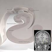
Hippocampal anatomy (illustration)
Case 3
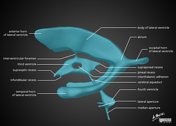
Jugular foramen (illustration)
Case 4
Middle ear anatomy - annotated CT
Case 5
Substantia nigra anatomy
Case 6
Alar and cruciform ligament anatomy
Case 7
Brain ventricle anatomy (illustration)
Case 8
Facial nerve anatomy - labeled CT
Case 9
Middle ear anatomy - annotated CT
Case 10
Neuroanatomy: superior cortex (diagrams)
Case 11
Subcortical U-fibers and juxtacortical lesions (illustration)
Case 12
Hippocampal anatomy (illustration)
Case 13
Veins of Trolard and Rosenthal
Case 14
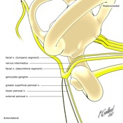
Geniculate ganglion (illustration)
Case 15
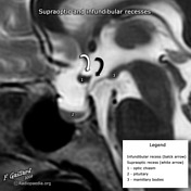
Supraoptic and infundibular recesses
Case 16
Angular gyrus and supramarginal gyrus (diagram)
Case 17
Basal vein of Rosenthal (annotated image)
Case 18
Frontal horn, intercaudate and inner table ratios (diagram)
Case 19
Anatomy: sulci of the brain
Case 20
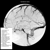
Cerebral veins (diagram)
Case 21
Midbrain anatomy
Case 22
Orbital anatomy (illustration)
Case 23
Posterior fossa vascular territories (illustration)
Case 24
Nerves of the internal acoustic meatus (diagram)
Case 25
Trigeminal nerve (normal)
Case 26
Cavernous sinus (diagram)
Case 27
Ossicles (illustration)
Case 28
Cerebellar tonsillar position (illustration)
Case 29
Layers of the scalp and meninges (illustrations)
Case 30
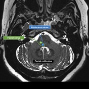
Abducens and facial cranial nerves and nuclei
Case 31
Arnold's and Jacobson's nerves (diagram)
Case 32
Normal MR spectroscopy (MRS)
Case 33
Middle cerebral artery branches
Case 34
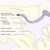
Pineal region anatomy (illustration)
Case 35
Left temporal bone (illustration)
Case 36
Cerebral veins (annotated DSA)
Case 37
Internal cerebral vein (annotated DSA)
Case 38
AC-PC line (diagram)
Case 39
Dural venous sinuses (illustration)
Case 40
Parieto-occipital region (diagram)
Case 41
Corpus callosum (annotated)
Case 42
Neuroanatomy - septal area (diagram)
Case 43
Neuroanatomy: insular cortex (diagrams)
Case 44
Cranial nerve nuclei (illustration) - deprecated
Case 45
Subcortical U-fibers and juxtacortical lesions (illustration)
Case 46
Midbrain anatomy
Case 47
CT facial bones/orbits sagittal - labeling questions
Case 48
CT head axial - labeling questions
Case 49
Fissula ante fenestram
Case 50
Post partum enlargement of pituitary
Case 51
Abducens nerve palsy
Case 52
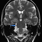
Demyelination with bilateral facial colliculus involvement
Case 53
Normal cranial nerves
Case 54
Polymicrogyria and absent septum pellucidum
Case 55
Normal petrous temporal bone axial CT - with labels
Case 56
Fibrous dysplasia - skull base
Case 57
Spinal cord lesion distribution
Case 58
Optic tract syndrome / infarct
Case 59
Hepatocellular carcinoma metastasis to meningioma (tumor-to-tumor metastasis)
Case 60
Oculomotor nerve palsy
Case 61
Scalp hematoma types (diagram)
Case 62
Brachial plexus (diagram)
Case 63
Brachial plexus anatomy
Case 64
Brachial plexus (normal)
Case 65
Temporal lobe atrophy post herpes simplex encephalitis
Case 66
Aicardi syndrome