UQ Med Yr 1 Chest: Lungs and pleura
Playlist contributed by
Craig Hacking
◉
◈
Play
Share
Playlist
Full screen playlist
Playlist with hidden diagnosis
Full screen playlist with hidden diagnosis
Playlist information
Playlist created:
22 Jul 2018 by
Craig Hacking
◉
◈
Last edited:
10 Dec 2019
Number of cases:
56
Number of slides:
21
rID:
119501
Systems:
Breast
,
Cardiac
,
Chest
,
Gastrointestinal
,
Head & Neck
,
Hepatobiliary
,
Interventional
,
Musculoskeletal
,
Oncology
,
Paediatrics
,
Spine
,
Trauma
,
Urogenital
,
Vascular
Visibility:
public
Show case titles
Slide 1
UQ_Med_Yr_1_Sem_1_-_Chest_Lungs_and_pleura.001.jpeg
Slide 2
UQ_Med_Yr_1_Sem_1_-_Chest_Lungs_and_pleura.001.jpeg
Slide 3
UQ_Med_Yr_1_Sem_1_-_Chest_Lungs_and_pleura.003.jpeg
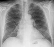
Case 4
Normal chest radiograph
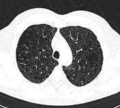
Case 5
Normal CT chest
Case 6
Normal chest x-ray - lobes (illustration)
Case 7
Bronchopulmonary segments (annotated CT)
Slide 8
UQ_Med_Yr_1_Sem_1_-_Chest_Lungs_and_pleura.001.jpeg
Slide 9
UQ_Med_Yr_1_Sem_1_-_Chest_Lungs_and_pleura.002.jpeg
Slide 10
air_bronch.004.jpg
Slide 11
air_bronch.005.jpg
Slide 12
air_bronch.006.jpg
Slide 13
sydney.001.jpeg
Case 14
Cardiomediastinal anatomy on chest radiography (annotated images)
Case 15
Left upper lobe consolidation due to pneumonia
Case 16
Left lower lobe pneumonia
Case 17
Right upper lobe consolidation: pediatric
Case 18
Silhouette sign of Felson - right middle lobe pneumonia
Case 19
Pneumonia - right lower lobe
Case 20
Right lower lobe consolidation - pneumonia
Case 21
Right lower lobe pneumonia
Case 22
Superior segment right lower lobe pneumonia
Slide 23
emerg_slides.001.jpeg
Case 24
Left lower lobe collapse
Case 25
Left lower lobe collapse
Case 26
Right upper lobe collapse
Case 27
Right lower lobe collapse and consolidation
Case 28
Left upper lobe collapse
Case 29
Right middle lobe collapse
Case 30
Endobronchial intubation
Slide 31
UQ_Med_Yr_1_Sem_1_-_Chest_Lungs_and_pleura.002.jpeg
Case 32
Solitary pulmonary nodule: melanoma metastasis
Case 33
Nipple markers: solitary pulmonary nodule
Case 34
Miliary tuberculosis
Case 35
Healed varicella pneumonia - miliary opacities
Case 36
Papillary thyroid carcinoma - with miliary metastases
Slide 37
UQ_Med_Yr_1_Sem_1_-_Chest_Lungs_and_pleura.003.jpeg
Case 38
Cardiac failure with Kerley B lines
Case 39
Cardiogenic pulmonary edema
Case 40
Acute pulmonary edema on CT
Slide 41
UQ_Med_Yr_1_Sem_1_-_Chest_Lungs_and_pleura.004.jpeg
Case 42
Severe COPD and pectus excavatum
Case 43
Emphysema
Case 44
Emphysema (diagrams)
Case 45
Centrilobular pulmonary emphysema
Case 46
Paraseptal emphysema
Case 47
Paraseptal emphysema and subpleural bullae
Case 48
Pan-lobular emphysema due to alpha-1-antitrypsin deficiency
Case 49
Usual interstitial pneumonia type pattern
Case 50
Asbestosis
Slide 51
UQ_Med_Yr_1_Sem_1_-_Chest_Lungs_and_pleura.005.jpeg
Case 52
Small right pleural effusion
Case 53
Hemothorax due to rib fractures
Case 54
Empyema in tension
Case 55
Pleural empyema
Case 56
Right hemithorax white-out: pleural effusion
Case 57
Spontaneous hemopneumothorax
Case 58
Pneumothorax
Case 59
Pneumothorax
Case 60
Pneumothorax on expiratory radiograph
Case 61
Tension pneumothorax
Case 62
Skin fold mimicking pneumothorax
Case 63
Pneumomediastinum and differential diagnoses
Case 64
Mach effect (diagram)
Case 65
Calcified pleural plaques
Case 66
Pleural thickening: illustrations
Case 67
Pleural thickening
Case 68
Post hemothorax pleural thickening and calcification
Slide 69
UQ_Med_Yr_1_Sem_1_-_Chest_Lungs_and_pleura.006.jpeg
Case 70
Epithelioid mesothelioma
Slide 71
Presentation_template.001.png
Case 72
Mesothelioma
Slide 73
UQ_Med_Yr_1_Sem_1_-_Chest_Lungs_and_pleura.007.jpeg
Slide 74
Presentation_template.002.png
Slide 75
Presentation_template.003.png