Neuro CT for nurses
Playlist contributed by
Arianne Churchill
Play
Share
Playlist
Full screen playlist
Playlist with hidden diagnosis
Full screen playlist with hidden diagnosis
Playlist information
Playlist created:
16 Oct 2019 by
Arianne Churchill
Last edited:
22 May 2020
Number of cases:
29
Number of slides:
16
rID:
125693
Systems:
Central Nervous System
,
Forensic
,
Head & Neck
,
Oncology
,
Paediatrics
,
Spine
,
Trauma
,
Vascular
Visibility:
public
Show case titles
Case 1
Normal brain CT
Case 2
Elderly CT brain
Slide 3
UQ_Med_Yr_1_Sem_2_-_Neuro_Brain_trauma_and_CVA.006.jpeg
Slide 4
brain.005.jpeg
Case 5
Extradural hematoma
Case 6
Extradural hematoma - venous
Slide 7
finale.001.jpeg
Case 8
Subdural hematoma, uncal herniation and Duret brainstem hemorrhage
Case 9
Subtle subdural hemorrhage and frontal contusion in trauma patient
Slide 10
bris.001.jpeg
Slide 11
bris.001.jpeg
Slide 12
bris.002.jpeg
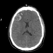
Case 13
Subdural hemorrhage - acute on warfarin
Case 14
Cerebral vascular territories (illustration)
Case 15
Subarachnoid hemorrhage due to aneurysm rupture
Case 16
Subarachnoid hemorrhage
Case 17
Subarachnoid hemorrhage
Case 18
Trauma and intraparenchymal cerebral hemorrhage
Slide 19
bris.003.jpeg
Case 20
Coup-contrecoup injury
Case 21
Inferior frontal lobe traumatic contusion
Slide 22
brain.001.jpeg
Case 23
Diffuse axonal injury and extra-axial bleed
Slide 24
brain.001.jpeg
Slide 25
brain.001.jpeg
Slide 26
brain.001.jpeg
Case 27
Subdural hemorrhage - on warfarin
Case 28
Subdural hematoma, uncal herniation and Duret brainstem hemorrhage
Case 29
Posterior cerebral artery territory infarct due to subdural hematoma and uncal herniation
Case 30
Uncal herniation with Kernohan phenomenon
Case 31
Subdural hemorrhage - on warfarin
Case 32
Extradural hematoma
Case 33
Falx subdural hematoma and sulcal subarachnoid hemorrhage
Slide 34
oedema_1.jpeg
Slide 35
oedema_2.jpeg
Case 36
Global hypoxic brain injury
Case 37
Vascular territories of the lateral cerebral cortex (illustration)
Case 38
Cerebral vascular territories in the midline (illustration)
Slide 39
common-variants-of-the-circle-of-willis-diagrams-1.jpg
Case 40
Motor and sensory homunculus (illustrations)
Slide 41
Presentation_template.003.png
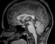
Case 42
Sagittal midline of the brain - normal anatomy
Case 43
Obstructive hydrocephalus
Case 44
Diffuse leptomeningeal glioneuronal tumor