VGHTC 201506
Playlist contributed by
Fu, Chien-hua
Play
Share
Playlist
Full screen playlist
Playlist with hidden diagnosis
Full screen playlist with hidden diagnosis
Playlist information
Playlist created:
23 Jun 2015 by
Fu, Chien-hua
Last edited:
30 May 2016
Number of cases:
155
Number of slides:
0
rID:
120001
Systems:
Central Nervous System
,
Chest
,
Forensic
,
Gastrointestinal
,
Head & Neck
,
Interventional
,
Musculoskeletal
,
Oncology
,
Paediatrics
,
Spine
,
Trauma
,
Urogenital
,
Vascular
,
Not Applicable
Visibility:
public
Show case titles
Case 1
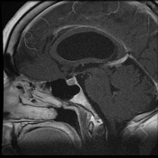
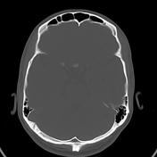
Fibrous dysplasia - skull base
Case 2
Cerebral arteriovenous malformation
Case 3
Band heterotopia
Case 4
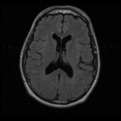
Alzheimeir's disease - probable
Case 5
Acoustic schwannoma - intracanalicular
Case 6
Towne view (skull AP axial view)
Case 7
White cerebellum sign of hypoxic-ischemic brain injury
Case 8
Cerebral venous thrombosis
Case 9
Glioblastoma IDH wild-type (with dural tail)
Case 10
Skull lateral view
Case 11
Third ventricle recesses
Case 12
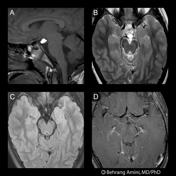
Ependymoma - MRS
Case 13
Cerebral arteriovenous malformation
Case 14
Venous sinus thrombosis with venous hemorrhage
Case 15
Multiple sclerosis
Case 16
Terminal internal carotid artery berry aneurysm causing acute 3rd cranial nerve palsy
Case 17
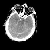
Normal CT head (cerebral and bone windows)
Case 18
Melanoma metastases - brain and spine
Case 19
Planum sphenoidale meningioma
Case 20
Cerebellar hemorrhage
Case 21
Solitary fibrous tumor (hemangiopericytoma)
Case 22
CNS lymphoma
Case 23
Secondary CNS lymphoma
Case 24
Clival and olfactory groove meningiomas
Case 25
Meningioma with rhabdoid component
Case 26
Normal pressure hydrocephalus
Case 27
Neurofibromatosis type 1
Case 28
Subdural hematoma - swirl sign
Case 29
Fazekas scale for white matter lesions
Case 30
Magnetic resonance parkinsonism index
Case 31
Dysembryoplastic neuroepithelial tumor (DNET) of frontal lobe
Case 32
Artery of Percheron infarct
Case 33
Subacute cerebral infarction - with MR spectroscopy
Case 34
Intracranial melanoma metastases
Case 35
Mesial temporal sclerosis
Case 36
Pilocytic astrocytoma
Case 37
Bifrontal leucotomy
Case 38
Clival chordoma
Case 39
Hemorrhagic cerebral metastasis
Case 40
Sinonasal adenocarcinoma with later dural involvement
Case 41
Cerebral metastasis - lung cancer
Case 42
Gliosarcoma
Case 43
Diffuse low grade astrocytoma
Case 44
Optic nerve sheath meningioma
Case 45
Petroclival meningioma
Case 46
Central pontine myelinolysis
Case 47
Occipital infarct
Case 48
Small polar encephalocele - temporal lobe
Case 49
Subretinal hemorrhage
Case 50
Corpus callosum agenesis
Case 51
Cavernous malformation (cavernous angioma or cavernoma)
Case 52
Crooke’s cell adenoma
Case 53
Multiple cerebral contusions and temporal bone fracture
Case 54
Low grade glioma (MR spectroscopy)
Case 55
Trigeminal schwannoma
Case 56
Left posterior cerebral artery territory stroke
Case 57
Glioblastoma NOS (multicentric)
Case 58
Intracranial hypotension - post spinal puncture
Case 59
Tuberous sclerosis with subependymal giant cell astrocytoma
Case 60
Blake's pouch cyst
Case 61
Calcified teflon granuloma
Case 62
Hypoxic-ischemic brain injury
Case 63
Intraventricular hemorrhage
Case 64
Encephalomalacia
Case 65
Left middle cerebral artery branch embolus causing stroke
Case 66
Thalamic lacunar infarct
Case 67
Meningioma - cerebellopontine angle
Case 68
Low grade glioma with radiation necrosis
Case 69
Adult intracranial hemorrhage on ultrasound
Case 70
Toxoplasmosis
Case 71
Terson syndrome
Case 72
Fahr disease
Case 73
Meningioma - histopathology
Case 74
Rathke cleft cyst - histopathology
Case 75
Copper beaten (photo)
Case 76
Tentorial angle
Case 77
Anterior communicating artery
Case 78
Marchiafava-Bignami disease
Case 79
Suprasellar cistern lipoma
Case 80
Cobblestones (photo)
Case 81
Pleomorphic xanthoastroctyoma (histology)
Case 82
Pleomorphic xanthoastrocytoma
Case 83
Intracranial ventricles - Gray's anatomy illustration
Case 84
Pituitary region (illustration)
Case 85
Blueberry (photo)
Case 86
Recurrent colloid cyst
Case 87
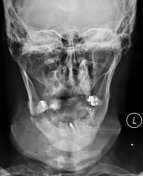
Bilateral mandibular fractures
Case 88
Sphenochoanal polyp
Case 89
Calvarial osteoma
Case 90
Persistent hyperplastic primary vitreous
Case 91
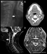
Calcific tendinitis of the longus colli muscle
Case 92
Pterygopalatine fossa
Case 93
Temporomandibular joint MRI anatomy
Case 94
Tram track (photo)
Case 95
External acoustic meatus (normal)
Case 96
Clival bone marrow signal
Case 97
Cochlear implant (illustration)
Case 98
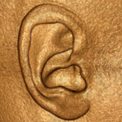
External ear
Case 99
Larynx (illustration)
Case 100
Malleus (illustration)
Case 101
Oval window and stapes
Case 102
Chagoma (photograph)
Case 103
Bony orbit (photo)
Case 104
Incisive canal cyst
Case 105
Primary uveal melanoma
Case 106
Skull AP view
Case 107
Skull PA view
Case 108
Inverted papilloma
Case 109
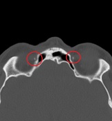
Normal petrous temporal bone axial CT - with labels
Case 110
Carotidynia
Case 111
Barium swallow aspiration
Case 112
Modified Stenvers view
Case 113
Nasal bone fracture
Case 114
Maxillary molar anatomy - illustration
Case 115
Calcified trochlear apparatus
Case 116
Orbital lymphoma
Case 117
Maxillary sinus - illustration
Case 118
Occipital spur
Case 119
Chronic brachial plexopathy and axillary artery occlusion post shoulder trauma
Case 120
Modic-type endplate changes: diagram
Case 121
Extravasation of cement into paravertebral veins in percutaneous vertebroplasty
Case 122
Atlanto-axial subluxation
Case 123
Chance fracture
Case 124
Traumatic adrenal hemorrhage
Case 125
Rugger jersey spine
Case 126
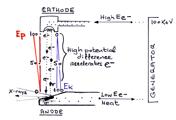
X-ray tube diagram
Case 127
Intervertebral disc disease nomenclature (illustration)
Case 128
Vertebral column CT anatomy (annotated image)
Case 129
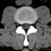
Disc herniation nomenclature - axial (annotated image)
Case 130
Sagittal localization of disc disease (annotated image)
Case 131
Spinal cord meninges (illustration)
Case 132
Jefferson fracture
Case 133
Facet joint injection - Scotty dog
Case 134
Spondylodiscitis
Case 135
Pott disease
Case 136
Spinal arteriovenous malformation
Case 137
Hangman fracture
Case 138
Cervical canal stenosis
Case 139
Ganglioneurocytoma
Case 140
Toothpaste (photo)
Case 141
Rugger-jersey spine
Case 142
Epidural lipomatosis
Case 143
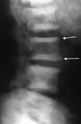
Schmorl node
Case 144
Schmorl nodes (discogram)
Case 145
Sugarcoated spine
Case 146
Block vertebra
Case 147
Sacral dimple
Case 148
Ankylosing spondylitis
Case 149
Spinal synovial cyst
Case 150
Normal cervical and thoracic spine MRI
Case 151
Fracture dislocation of cervical facet joint
Case 152
Vertebral and epidural metastases
Case 153
Epidural abscess and facet joint septic arthritis
Case 154
Spondylolysis and spondylolisthesis
Case 155
Spondylodiscitis and epidural abscess caused by esophageal perforation due to a metallic stent