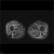
MRI findings in muscle pathology can be split into one of three broad categories:
- oedema, e.g. trauma, infection, inflammation
- fatty deposition, e.g. atrophy
- mass, e.g. haematoma, abscess, tumour, vascular malformation
The functional unit in skeletal muscle is the muscle fibre which is surrounded by a thin layer of connective tissue, the endomysium, and a network of capillaries.
20–80 muscle fibres are grouped together to form a fascicle which is surrounded by perimysium. Larger vessels run between the fascicles. Each muscle is formed by a collection of fascicles surrounded by a thick connective tissue layer, the epimysium, that condenses at the end of the muscles to form tendons.
Muscle oedema is one of three (oedema, atrophy, mass) ways in which muscle pathology presents. Oedema may be the result of a range of traumatic, infective and inflammatory conditions and a number of examples have been given here.


 Unable to process the form. Check for errors and try again.