


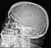


Skull solitary lesions:
Inspect the margins:
- permeative margins suggest aggressive lesion such as mets or infection
- dense sclerotic margins suggest slow growing lesions such as epidermoids and dermoids
Inspect internal structure:
- central residual bone density is classic for EG but can be seen in mets/osteomyelitis and epidermoid
- spoke wheel or reticulated internal pattern is classic for haemangioma
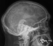
Age:
- children lytic skull lesion > EG, less likely neuroblastoma mets or leukemia
- children and young adults > haemangioma
- adults and elderly > mets most common
DDx for lytic/sclerotic lesions:
Haemangioma
Epidermoid/dermoid
Lymphoma/leukemia/leptomenigneal cyst
Paget disease/post surgical
Metastases/multiple myeloma (plasmacytoma)
Eosinophilic granumal/encephalocele


 Unable to process the form. Check for errors and try again.