Classifications / Diagrams
Playlist contributed by
Blake Milton
Play
Share
Playlist
Full screen playlist
Playlist with hidden diagnosis
Full screen playlist with hidden diagnosis
Playlist information
Playlist created:
12 Feb 2023 by
Blake Milton
Last edited:
13 Feb 2023
Number of cases:
125
Number of slides:
0
rID:
162519
Systems:
Cardiac
,
Central Nervous System
,
Chest
,
Forensic
,
Gastrointestinal
,
Gynaecology
,
Haematology
,
Head & Neck
,
Hepatobiliary
,
Musculoskeletal
,
Obstetrics
,
Oncology
,
Paediatrics
,
Spine
,
Trauma
,
Urogenital
,
Vascular
,
Not Applicable
Visibility:
public
Show case titles
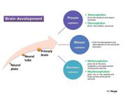
Case 1
Brain development (diagram)
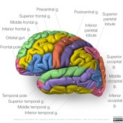
Case 2
Neuroanatomy: lateral cortex (diagrams)
Case 3
Brainstem arterial territories (diagrams)
Case 4
Subcortical U-fibers and juxtacortical lesions (illustration)
Case 5
Brain venous vascular territories (diagram)
Case 6
Diffuse glioma classification (WHO 5th Edition, 2021)
Case 7
Scalp hematoma types (diagram)
Case 8
Germinal matrix hemorrhage grading
Case 9
Brain herniation types
Case 10
Diagram - intracranial hemorrhage
Case 11
Subdural hygroma illustration
Case 12
Nerves of the internal acoustic meatus (diagram)
Case 13
Pterygopalatine fossa (diagram)
Case 14
Internal carotid artery segments (illustration)
Case 15
Chandler classification of orbital infections (diagram)
Case 16
Orbital apex (diagram)
Case 17
Le Fort fracture classification (illustration)
Case 18
Nasal surface anatomy (diagram)
Case 19
ACR TI-RADS scoring system (diagram)
Case 20
Pharyngeal arches (creative commons diagram)
Case 21
Thyroglossal duct (diagram)
Case 22
Disc herniation nomenclature
Case 23
Modic-type endplate changes: diagram
Case 24
Levine and Edwards classification of hangman fractures (diagrams)
Case 25
Gehweiler classification of atlas fractures (diagrams)
Case 26
Cervical facet dislocation diagrams
Case 27
Odontoid fracture classification (diagram)
Case 28
AOSpine Injury Classifications
Case 29
Lymph node regions (illustration)
Case 30
Bronchopulmonary segments (annotated CT)
Case 31
Thoracic lymph node stations (annotated CT)
Case 32
Classification of pulmonary sequestration
Case 33
Pulmonary nodule patterns (diagram)
Case 34
Classification of pneumonia
Case 35
Emphysema (diagrams)
Case 36
Krichenko angiographic classification of patent ductus arteriosus (illustration)
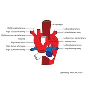
Case 37
Double aortic arch (diagram)
Case 38
Double aortic arch (diagram)
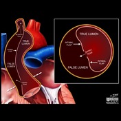
Case 39
Aortic dissection DeBakey classification (illustration)
Case 40
Pathogenesis of aortic dissection (illustration)
Case 41
Pathogenesis of aortic intramural hematoma (illustration)
Case 42
Pathogenesis of penetrating atherosclerotic ulcer (illustration)
Case 43
Endoleak classification (diagram)
Case 44
Retroperitoneal spaces: diagram
Case 45
Pyloric stenosis - diagram
Case 46
Couinaud classification (diagram)
Case 47
Pancreatic duct anatomic variation (diagram)
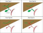
Case 48
Kasai classification of biliary atresia (diagram)
Case 49
Choledochal cyst types (diagram)
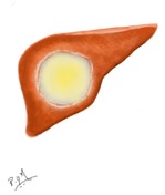
Case 50
Hydatid disease of the liver (illustrations)
Case 51
Liver trauma grading (diagrams)
Case 52
Barcelona clinic liver cancer (BCLC) staging classification
Case 53
Pancreatic trauma grading (diagrams)
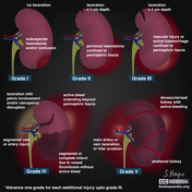
Case 54
Renal trauma grading diagrams
Case 55
Bosniak classification of renal cysts (illustrations)
Case 56
Diagram - circumcaval ureter
Case 57
Renal trauma grading (diagrams)
Case 58
Crossed fused renal ectopia (diagram)
Case 59
PI-RADS flowchart
Case 60
Rectus sheath (diagram)
Case 61
Inguinal canal (diagram)
Case 62
Maydl's hernia (diagram)
Case 63
Bell clapper deformity (diagram)
Case 64
Uterine anatomical abnormalities (illustrations)
Case 65
Uterine version and flexion (diagrams)
Case 66
Uterine leiomyoma (fibroid) classification system (illustration)
Case 67
Endometriosis (diagrams)
Case 68
Ectopic pregnancy distribution (diagram)
Case 69
Variation in placental morphology: diagrams
Case 70
Variation in placental adherence: diagrams
Case 71
Twin reversed arterial perfusion - illustration
Case 72
Salter-Harris illustrations
Case 73
Periosteal reactions (diagram)
Case 74
Rockwood classification of acromioclavicular joint injury
Case 75
Illustration - common labral injuries
Case 76
Cubital fossa (diagram)
Case 77
AO/OTA classification of distal humeral fractures
Case 78
Supracondylar fracture (illustrations)
Case 79
Wrist extensor compartments (diagram)
Case 80
Carpal tunnel (diagram)
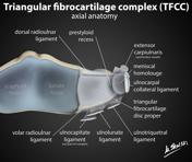
Case 81
Triangular fibrocartilage complex (TFCC) anatomy illustrations
Case 82
Atzei classification of peripheral TFCC tears
Case 83
Scaphoid fractures - Mayo classification
Case 84
Dorsal and volar intercalated segmental instability and normal carpal bone anatomy (illustrations)
Case 85
Progressive perilunate instability (diagram)
Case 86
Hand arthropathies - distribution (diagram)
Case 87
Thumb metacarpal fractures (illustration)
Case 88
Diagram - distribution of solitary enchondromas of the hand
Case 89
Femoral triangle (diagram)
Case 90
Normal marrow conversion (diagram)
Case 91
Acetabular angle - diagram
Case 92
Coxa vara and coxa valga (diagram)
Case 93
Graf Hip Angles - Alpha and Beta (illustration)
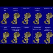
Case 94
Judet-Letournel classification of acetabular fractures
Case 95
Legg-Calve-Perthes disease: Salter-Thompson classification
Case 96
Garden classification of hip fractures (diagram)
Case 97
Popliteal fossa (diagram)
Case 98
Pes anserinus tendon (diagram)
Case 99
Bone lesion differential diagnosis - illustrations
Case 100
Distribution of unicameral bone cysts (diagram)
Case 101
Distribution of aneurysmal bone cysts (diagram)
Case 102
Meniscus ultrastructure diagram
Case 103
Meniscal tear types - diagram
Case 104
MRI grading system for meniscal signal intensity (diagrams)
Case 105
Synovial osteochondromatosis (illustration)
Case 106
Diffuse tenosynovial giant cell tumor (illustration)
Case 107
Lipoma arborescens (illustration)
Case 108
Classification of ACL avulsion fractures (diagram)
Case 109
Schatzker classification of tibial plateau fractures (illustrations)
Case 110
Kfuri and Schatzker classification of tibial plateau fractures
Case 111
Chondromalacia grading
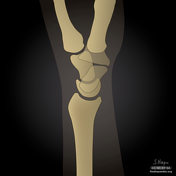
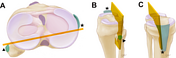
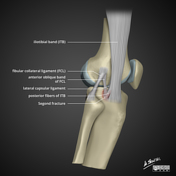
Case 112
Segond fracture (diagram)
Case 113
Diagram - Metaphyseal diaphyseal angle of Drennan
Case 114
Illustration for lateral patellar dislocation
Case 115
Ankle and foot ligaments (Gray's illustrations)
Case 116
Weber fracture classification (illustration)
Case 117
Weber classification of ankle fractures
Case 118
Sanders classification of calcaneal fractures
Case 119
Os naviculare: diagrams
Case 120
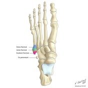
Fractures of the proximal 5th metatarsal
Case 121
Nunley-Vertullo classification of Lisfranc injuries (illustrations)
Case 122
Hounsfield scale (diagram)
Case 123
Evolution of MRI signal characteristics of intracranial hemorrhage (diagram)
Case 124
Evolution of CT density of intracranial hemorrhage (diagram)
Case 125
Timeline diagram of MRI and CT characteristics of intracerebral hemorrhage