RANZCR Part 2 Revision
Playlist contributed by
Namit Mathur
Play
Share
Playlist
Full screen playlist
Playlist with hidden diagnosis
Full screen playlist with hidden diagnosis
Playlist information
Playlist created:
8 Sep 2023 by
Namit Mathur
Last edited:
5 May 2024
Number of cases:
58
Number of slides:
0
rID:
175085
Systems:
Cardiac
,
Central Nervous System
,
Chest
,
Gastrointestinal
,
Gynaecology
,
Haematology
,
Head & Neck
,
Musculoskeletal
,
Obstetrics
,
Oncology
,
Paediatrics
,
Spine
,
Trauma
,
Urogenital
,
Vascular
Visibility:
public
Show case titles
Case 1
Evolution of MRI signal characteristics of intracranial haemorrhage (diagram)
Case 2
Thumb metacarpal fractures (illustration)
Case 3
Hand arthropathies - distribution (diagram)
Case 4
Cystic lung diseases (illustrations)
Case 5
Bone lesion differential diagnosis - illustrations
Case 6
Chronic vs acute osteomyelitis (illustration)
Case 7
Paediatric posterior fossa tumours illustration
Case 8
Cerebral amyloid angiopathy (illustration)
Case 9
Pleural thickening: illustrations
Case 10
Emphysema (diagrams)
Case 11
Pulmonary nodule patterns (diagram)
Case 12
Cervical ankylosis - DISH, OPLL, AS - illustrations
Case 13
Illustration of scurvy signs
Case 14
Mallet finger (illustration)
Case 15
Salivary gland tumour subtypes (table)
Case 16
Diffuse glioma classification (WHO 5th Edition, 2021)
Case 17
Spinal cord lesion distribution
Case 18
Uterine anatomical abnormalities (illustrations)
Case 19
Disc herniation nomenclature
Case 20
Pyloric stenosis - diagram
Case 21
Illustration - common labral injuries
Case 22
Disc herniation nomenclature
Case 23
Patterns of knee injury
Case 24
Scapholunate advanced collapse (illustration)
Case 25
Scaphoid non-union advanced collapse (illustration)
Case 26
Normal wrist alignment, dorsal and volar intercalated segmental instability (illustration)
Case 27
Uterine leiomyoma (fibroid) classification system (illustration)
Case 28
Incomplete spinal cord syndromes (illustrations)
Case 29
Cerebellar tonsillar position (illustration)
Case 30
Variation in cord insertion
Case 31
Otic capsule: annotated CT
Case 32
CT neck with annotated scrollable images
Case 33
Brain venous vascular territories (diagram)
Case 34
Le Fort fracture classification (illustration)
Case 35
Morphological types of bronchiectasis (illustration)
Case 36
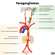
Paragangliomas (illustration)
Case 37
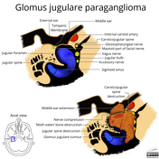
Jugular paraganglioma (axial view illustration)
Case 38
Periosteal reaction types
Case 39
Lymph node levels (illustration)
Case 40
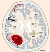
Germinal matrix haemorrhage grading
Case 41
Syndesmophyte v Osteophyte (illustration)
Case 42
Bosniak classification of renal cysts (illustrations)
Case 43
Pelvic apophyseal avulsion fractures (annotated image)
Case 44
Carpal dislocations (illustrations)
Case 45
Lipohaemarthrosis and haemarthrosis (illustrations)
Case 46
Sinus tarsi (diagram)
Case 47
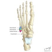
Fractures of the proximal 5th metatarsal
Case 48
Elbow ossification centres
Case 49
Placenta praevia spectrum
Case 50
Ectopic pregnancy distribution (diagram)
Case 51
Slipped capital femoral epiphysis (illustrations)
Case 52
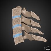
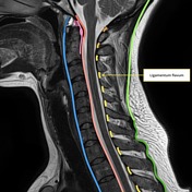
Ligaments of the cervical spine (annotated image)
Case 53
Sickle cell disease with osseous changes and splenomegaly
Case 54
Knee radiograph checklist (illustration)
Case 55
Mitral heart
Case 56
Placenta accreta spectrum (illustration)
Case 57
Long-term epilepsy-associated tumours (LEAT)
Case 58
Temporal bone fracture - diagram