Diagram
Playlist contributed by
Krzysztof Dud
Play
Share
Playlist
Full screen playlist
Playlist with hidden diagnosis
Full screen playlist with hidden diagnosis
Playlist information
Playlist created:
22 Jun 2024 by
Krzysztof Dud
Last edited:
22 Jun 2024
Number of cases:
65
Number of slides:
0
rID:
191490
Systems:
Cardiac
,
Central Nervous System
,
Chest
,
Forensic
,
Gastrointestinal
,
Gynaecology
,
Head & Neck
,
Hepatobiliary
,
Musculoskeletal
,
Obstetrics
,
Oncology
,
Paediatrics
,
Spine
,
Trauma
,
Urogenital
,
Vascular
,
Not Applicable
Visibility:
public
Show case titles
Case 1
Biliary duct anatomic variation (diagram)
Case 2
Deep spaces of the head and neck - annotated MRI
Case 3
Brain venous vascular territories (diagram)
Case 4
Cerebral vascular territories (illustration)
Case 5
Cerebral perfusion parameters
Case 6
Chondromalacia grading
Case 7
Cranial nerve nuclei (axial diagrams)
Case 8
Coxa vara and coxa valga (diagram)
Case 9
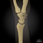
Dorsal and volar intercalated segmental instability and normal carpal bone anatomy (illustrations)
Case 10
Distal radial fractures (illustration)
Case 11
Diagram - Bismuth-Corlette classification of perihilar cholangiocarcinoma
Case 12
Diagram - Bismuth-Corlette classification of perihilar cholangiocarcinoma
Case 13
Diagram - Bismuth-Corlette classification of perihilar cholangiocarcinoma
Case 14
Ectopic pregnancy distribution (diagram)
Case 15
Endoleak classification (diagram)
Case 16
Facet dislocation diagrams
Case 17
Gamekeeper's thumb and Stener lesion (illustrations)
Case 18
Late gadolinium enhancement patterns (diagram)
Case 19
Keros classification (illustration)
Case 20
Hounsfield scale (diagram)
Case 21
Hand arthropathies - distribution (diagram)
Case 22
Motor and sensory homunculus (illustrations)
Case 23
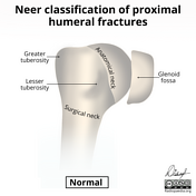
Neer classification of proximal humeral fractures
Case 24
Morel-Lavallée Illustrations
Case 25
Mesentero-axial gastric volvulus
Case 26
May-Thurner syndrome
Case 27
Lymph node regions (illustration)
Case 28
Lymph node levels (illustration)
Case 29
Lumbar spinal canal stenosis grading systems by Lee and Schizas
Case 30
Lumbar neuroforaminal stenosis - illustrations
Case 31
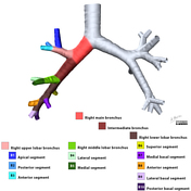
Lobar and segmental bronchial anatomy
Case 32
Liver trauma grading (diagrams)
Case 33
Lisfranc injury - Myerson classifications (illustrations)
Case 34
Lichtman classification of Kienböck disease (illustration)
Case 35
Levine and Edwards classification of hangman fractures (diagrams)
Case 36
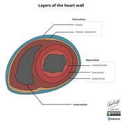
Left ventricular myocardial segmentation and coronary artery territories
Case 37
Radial inclination angle
Case 38
Pulled elbow syndrome (illustration)
Case 39
Pseudoaneurysm
Case 40
Prostate anatomy
Case 41
Progressive perilunate instability (diagram)
Case 42
PI-RADS v2.1 flowchart
Case 43
PI-RADS flowchart
Case 44
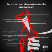
Persistent carotid-vertebrobasilar anastomoses
Case 45
Wrist extensor tendons (reporting aid)
Case 46
Wrist extensor compartments (diagram)
Case 47
Vascularization of the female reproductive system (Gray's illustration)
Case 48
Variant anatomy of the odontoid process of C2
Case 49
Urachus (illustration)
Case 50
Triangle of Guillain and Mollaret
Case 51
Timeline diagram of MRI and CT characteristics of intracerebral hemorrhage
Case 52
Spinal cord (illustrations)
Case 53
Skull base superior and inferior views (illustrations)
Case 54
Shoulder ultrasound - supraspinatus identification (illustration)
Case 55
Shoulder ultrasound - subscapularis identification illustration
Case 56
Shoulder ultrasound - infraspinatus identification illustration
Case 57
Sanders classification of calcaneal fractures
Case 58
Salter-Harris illustrations
Case 59
Salient features in different arthropathies (illustration)
Case 60
Sacral plexus (diagram)
Case 61
Rockwood classification of acromioclavicular joint injury
Case 62
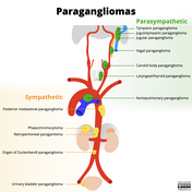
Paragangliomas (illustration)
Case 63
Pancreatic trauma grading (diagrams)
Case 64
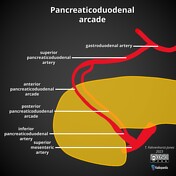
Pancreaticoduodenal arcade
Case 65
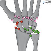
Ossicles of the wrist and hand (illustration)