


18 results found
Case
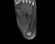
Baxter’s nerve impingement

Published
20 Jun 2019
95% complete
MRI
Case
Baxter’s nerve impingement

Published
03 May 2021
77% complete
MRI
Article
Inferior calcaneal nerve
The inferior calcaneal nerve (or Baxter nerve) is a mixed nerve in the foot.
Gross anatomy
Origin
Originates from the lateral plantar nerve at the level of the medial malleolus.
Course
Courses between the abductor hallucis muscle and the quadratus plantae and along the medial border of the...
Article
Baxter neuropathy
Baxter neuropathy is a nerve entrapment syndrome resulting from the compression of the inferior calcaneal nerve (Baxter nerve).
Clinical presentation
heel pain with maximal tenderness over the course of the inferior calcaneal nerve (on the plantar medial aspect of the foot and anterior to the ...
Article
Sciatic nerve motor distribution
The motor distribution of the sciatic nerve is as follows
Thigh
tibial division (posterior compartment of the thigh)
long head of biceps femoris
semimembranosus
semitendinosus
hamstring part of adductor magnus
peroneal division (superficial and lateral to tibial division)
short head of b...
Article
Nerve compression syndrome
Nerve compression syndromes or nerve entrapment neuropathies are a group of several nerve disorders associated with sensory and/or motor loss resulting from nerve compression.
Epidemiology
Nerve compression syndromes are common 1-5 and can account for 10-20% of cases in specialist clinics of n...
Case
Posterior tibial vein thrombosis causing tarsal tunnel syndrome

Published
29 Mar 2023
92% complete
MRI
Ultrasound
Annotated image
Article
Medial plantar nerve entrapment
Medial plantar nerve entrapment or compression syndrome, also known as jogger’s foot is a nerve compression syndrome of the medial plantar nerve either in the distal tarsal tunnel or beneath the plantar arch at the knot of Henry.
Epidemiology
Medial plantar nerve entrapment is a rather rare ty...
Article
Abductor hallucis muscle
The abductor hallucis muscle forms the medial margin of the foot and contributes to a soft tissue bulge on the medial side of the sole.
Summary
origin: medial process of calcaneal tuberosity
insertion: medial side of base of proximal phalanx of great toe
action: abducts and flexes great toe ...
Article
Foot pain
Foot pain is a very common symptom. The differential diagnosis depends mainly on age, weight, level of physical activity, and the exact location of the pain.
As neoplastic lesions are ubiquitary, they will not be added to the sections below.
Hindfoot pain
inferior heel pain
trauma...
Article
Abductor digiti minimi muscle (foot)
The abductor digiti minimi muscle is on the lateral side of the foot and contributes to the large lateral plantar eminence on the sole.
Summary
origin: lateral and medial processes of calcaneal tuberosity, and band of connective tissue connecting calcaneus with base of the fifth metatarsal
in...
Case
Plantar fasciitis, abductor digiti minimi atrophy and probable Baxter neuropathy

Published
02 Aug 2014
77% complete
X-ray
MRI
Case
Baxter neuropathy

Published
20 Nov 2013
78% complete
MRI
CT
Case
Baxter neuropathy

Published
02 Aug 2015
95% complete
MRI
X-ray
Case
Baxter neuropathy

Published
11 Dec 2018
77% complete
MRI
Case
Hypertrophic abductor hallucis muscle
Published
10 Feb 2016
92% complete
MRI
Article
MRI of the ankle (an approach)
MRI of the ankle is one of the more frequent examinations faced in daily radiological practice. This approach is an example of how to create a radiological report of an MRI of the ankle with coverage of the most common anatomical sites of possible pathology, within the ankle without claim for co...
Article
Aggressive vertebral hemangioma
Aggressive vertebral hemangiomata are a rare form of vertebral hemangiomata where significant vertebral expansion, extra-osseous component with epidural extension, disturbance of blood flow, and occasionally compression fractures can be present causing spinal cord and/or nerve root compression 1...


 Unable to process the form. Check for errors and try again.