


465 results found
Article
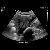
Ovarian cyst
Ovarian cysts are commonly encountered in gynecological imaging and vary widely in etiology from physiological to complex benign to neoplastic.
Pathology
Small cystic ovarian structures should be considered normal ovarian follicles unless the patient is pre-pubertal, post-menopausal, pregnant,...
Article
Ruptured ovarian cyst
Ruptured ovarian cysts are one of the most common causes of acute pelvic pain in premenopausal women. The sonographic appearance depends on whether a simple or hemorrhagic ovarian cyst ruptures, and whether the cyst has completely collapsed. The most important differential consideration is a rup...
Article
Hemorrhagic ovarian cyst
Hemorrhagic ovarian cysts usually result from hemorrhage into a corpus luteum or other functional cyst. Radiographic features are variable depending on the age of the hemorrhage. They typically resolve within 8 weeks.
Clinical presentation
Patients may present with sudden-onset pelvic pain, p...
Article
Functional ovarian cyst
A functional ovarian cyst is a term given to a group of non neoplastic type of ovarian cysts. A large proportion of ovarian cysts detected on imaging are functional ovarian cysts. Entities that fall under this group include
ovarian follicular cysts
corpus luteum cysts
theca lutein cysts
Func...
Article
Fetal ovarian cyst
Fetal ovarian cysts refer to an ovarian cyst detected antenatally in a female fetus. They are relatively uncommon and are usually diagnosed in the 3rd trimester 5.
Epidemiology
From autopsy studies, they are found in up to 30% of fetuses 1.
Pathology
The exact etiology is not well known at t...
Article
Ovarian follicular cyst
An ovarian follicular cyst is type of simple physiological ovarian cyst.
Terminology
The terms "ovarian cyst" and "ovarian follicular cyst" are often used interchangeably. These two terms describe lesions >3 cm, and it is important to differentiate them from an "ovarian follicle" which is <3 c...
Article
Tip of the iceberg sign (ovarian dermoid cyst)
Tip of the iceberg sign refers to one of the characteristic appearances of an ovarian dermoid cyst. If there are echogenic cyst contents of sebum and hair, they cause marked posterior acoustic attenuation so that only the superficial part of the cyst is seen. Just like an iceberg, you may only b...
Article
Dot dash pattern (ovarian dermoid cyst)
The dot-dash pattern (dermoid mesh) is one of the characteristic sonographic appearances of an ovarian dermoid cyst. It refers to the short and long echogenic lines which are often seen within a dermoid cyst and are due to the presence of hair.
Case
Ovarian cyst

Published
23 Apr 2010
92% complete
CT
Case
Ovarian cyst

Published
13 Apr 2016
89% complete
CT
Case
Ovarian cyst - prepubertal

Published
12 Jul 2023
95% complete
Pathology
X-ray
Ultrasound
CT
Case
Ovarian cyst torsion

Published
23 Apr 2023
95% complete
CT
Case
Giant ovarian cyst

Published
13 Sep 2023
95% complete
CT
Case
Hemorrhagic ovarian cyst
Published
16 Apr 2020
95% complete
Ultrasound
CT
Case
Hemorrhagic ovarian cyst

Published
16 May 2012
94% complete
Ultrasound
Pathology
Case
Ovarian cyst

Published
08 Dec 2013
79% complete
Ultrasound
Case
Ruptured ovarian cyst

Published
17 Jan 2024
89% complete
Ultrasound
CT
Case
Ovarian cyst

Published
27 Jul 2011
74% complete
CT
Ultrasound
Case
Follicular ovarian cyst

Published
22 Nov 2016
84% complete
CT
Ultrasound
MRI
Case
Hemorrhagic ovarian cyst

Published
13 Oct 2021
82% complete
Ultrasound


 Unable to process the form. Check for errors and try again.