


120 results found
Case
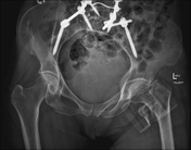
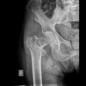
Acute pubic rami fractures

Published
29 Aug 2020
79% complete
X-ray
Article
Atypical femoral fracture
Atypical femoral fractures, also known as bisphosphonate-related proximal femoral fractures, are an example of insufficiency fractures, although the direct causative link remains somewhat controversial 2. The atypical fracture pattern occurs in the femur shaft and may be unilateral or bilateral....
Case
Avascular necrosis of the left hip

Published
01 Mar 2019
80% complete
MRI
Case
Bilateral femoral neck fractures

Published
02 Jul 2018
79% complete
X-ray
Case
Bladder hematoma

Published
11 May 2023
95% complete
Ultrasound
CT
Article
Cam morphology (femoroacetabular impingement)
Cam morphology refers to an abnormal morphology of the femoral head-neck junction interlinked with an osseous asphericity of the femoral head. It is one possible cause of femoroacetabular impingement (FAI).
Terminology
Cam morphology is also commonly referred to as 'cam deformity', 'cam lesion...
Case
CAM type femoroacetabular impingement with fracture

Published
03 Aug 2014
95% complete
X-ray
MRI
Article
Chondrosarcoma
Chondrosarcomas are a heterogeneous group of malignant cartilaginous tumors most commonly found in older patients. They can arise de novo or secondary from an existing benign cartilaginous neoplasm. On imaging, these tumors have ring-and-arc chondroid matrix mineralization with aggressive featur...
Case
Comminuted intertrochanteric neck of femur fracture (X-Ray and CT)
Published
25 Apr 2023
98% complete
X-ray
CT
Playlist
Core Conditions 03.2 - lower limb trauma pre-reading


18 cases
Welcome to The Royal Melbourne Hospital Core Conditions in Emergency Radiology. This module is focussed on lower limb trauma.The followin...
Article
Coxa vara
Coxa vara describes a hip deformity where the femoral neck-shaft angle is decreased, usually defined as <120°.
Pathology
It can be congenital or acquired. The common mechanism in congenital cases is a failure of the medial growth of the physeal plate 3.
Etiology
The etiology of coxa vara w...
Article
CT hip (protocol)
The CT hip protocol serves as an examination for the evaluation of the hip joint. It is often performed as a non-contrast study. However, it can be combined with a CT arthrogram for the evaluation of chondral and/or labral tears or a femoral neck version scan.
Note: This article aims to frame a...
Case
Cut-out lag screw, neck of femur fracture gamma nail

Published
03 Dec 2015
80% complete
CT
Article
Delbet classification
The Delbet classification helps predict the risk of avascular necrosis of the femoral head in neck of femur fractures, as well as determine operative vs non-operative management 1.
Classification
type I: trans-epiphyseal separation
fracture through proximal femoral physis, representing Salte...
Article
Dual-energy x-ray absorptiometry
Dual-energy x-ray absorptiometry (DEXA or DXA) is a technique used to aid in the diagnosis of osteopenia and osteoporosis.
Radiographic features
Values are calculated for the lumbar vertebrae and femur preferentially, and if one of those sites is not suitable (e.g. artifact, patient mobility)...
Case
Fallen fragment sign in neck of femur fracture

Published
23 May 2016
95% complete
CT
Article
Fatigue fracture
Fatigue fractures (also known as overuse fractures) are a type of stress fracture due to abnormal stresses on normal bone. They should not be confused with an insufficiency fracture, which occurs due to normal stresses on abnormal bone. Plain radiographs typically demonstrate a linear sclerotic ...
Case
Fatigue fracture - femoral neck

Published
22 Apr 2024
90% complete
MRI
Fluoroscopy
Case
Femoral head blood supply

Published
15 Apr 2022
47% complete
Diagram
Case
Femoral neck fracture
Published
05 Jul 2012
79% complete
X-ray


 Unable to process the form. Check for errors and try again.