
Search results for “twinkle artifact”


32 results found
Article
Twinkling artifact
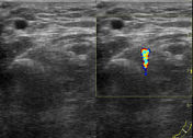
Twinkling artifact is seen with color flow Doppler ultrasound 1. It occurs as a focus of alternating colors on Doppler signal behind a reflective object (such as a calculus or air), which gives the appearance of turbulent blood flow 2. It appears with or without an associated color comet tail ar...
Case
Twinkling artifact of vesicoureteric junction calculus

Published
07 May 2020
94% complete
Ultrasound
Annotated image
Case
Twinkling artifact caused by kidney stone

Published
22 Nov 2020
92% complete
CT
Ultrasound
Case
Vesicoureteric junction calculus - value of twinkling artifact

Published
07 May 2020
91% complete
Ultrasound
Case
Twinkling artifact - renal stone

Published
04 Oct 2022
75% complete
Ultrasound
Case
Twinkle artifact of renal calculi

Published
28 Mar 2017
75% complete
Ultrasound
Case
Vesicoureteric junction calculus with twinkling artifact

Published
31 Jan 2015
69% complete
Ultrasound
Case
Vesicoureteric junction and renal calculi: the value of twinkle artifact

Published
04 Feb 2011
75% complete
Ultrasound
Case
Twinkling artifact caused by calcified renal cyst

Published
22 Nov 2018
68% complete
Ultrasound
Case
Twinkling and color comet tail artifacts

Published
23 Jun 2014
91% complete
Ultrasound
Article
Ultrasound artifacts
Ultrasound artifacts are commonly encountered and familiarity is necessary to avoid false diagnoses. In some cases, specific artifacts can even offer valuable diagnostic information. For instance, some artifacts may be indicative of certain pathologies. They are not to be confused with ultrasou...
Article
Color comet tail artifact
The color comet tail artifact is an ultrasonographic sign seen in a number of situations when color Doppler scanning is performed.
Typically the artifact, which resembles the grey scale comet tail artifact, is seen in a situation when a small highly reflective (usually calcific) object is inter...
Article
Pulse repetition frequency
Pulse repetition frequency (PRF) indicates the number of ultrasound pulses emitted by the transducer over a designated period of time. It is typically measured as pulses per second or hertz (Hz). In medical ultrasound the typically used range of PRF varies between 1 and 10 kHz 1. PRF is defined ...
Article
Astronomical inspired signs
Many signs in radiology have been inspired by astronomical phenomena:
comet tail (disambiguation)
comet tail artifact (ultrasound)
color comet tail artifact
comet tail sign (chest)
comet tail sign (phleboliths)
earth-heart sign
galaxy sign (chest)
loss of half-moon overlap sign
milky wa...
Article
Color flow Doppler (ultrasound)
The use of color flow Doppler (CFD) or color Doppler imaging (CDI) (or simply color Doppler) sonography allows the visualization of flow direction and velocity within a user defined area. A region of interest is defined by the sonographer, and the Doppler shifts of returning ultrasound waves wit...
Article
Choledocholithiasis
Choledocholithiasis denotes the presence of gallstones within the bile ducts (including the common hepatic duct/common bile duct).
Epidemiology
Choledocholithiasis is relatively common, seen in up to 20% of patients undergoing cholecystectomy for gallstone-related complaints 2.
Clinical prese...
Article
Urolithiasis
Urolithiasis refers to the presence of calculi anywhere along the course of the urinary tracts. For the purpose of the article, the terms urolithiasis, nephrolithiasis, and renal/kidney stones are used interchangeably, although some authors have slightly varying definitions of each.
See main a...
Article
Gallstones
Gallstones, also called cholelithiasis, are concretions that may occur anywhere within the biliary system, most commonly within the gallbladder.
Terminology
Gallstones (cholelithiasis) describe stone formation at any point along the biliary tree. Specific names can be given to gallstones depe...
Article
Ocular foreign body
An ocular foreign body occurs when an orbital foreign body intrudes into the globe itself, often threatening vision, and requiring urgent surgical removal.
Clinical presentation
Patients present in a highly variable manner based on the precise intraocular location and properties of the foreign...
Case
Foreign body - forearm
Published
30 Jun 2016
88% complete
Ultrasound


 Unable to process the form. Check for errors and try again.