
Items tagged “anatomy”
380 results found
Article
Maxillary vein
The maxillary vein drains the pterygoid plexus and joins with the superficial temporal vein to form the retromandibular vein in the substance of the parotid gland 1.
Gross anatomy
The pterygoid plexus, and by extension, the maxillary vein helps to drain the areas supplied by the maxillary arte...
Case
Superficial lateral malleolus bursa

Published
20 Oct 2018
53% complete
MRI
Case
Sternocostal and interchondral joint anatomy (illustration)

Published
14 Feb 2019
22% complete
Diagram
Article
Interventricular septum
The interventricular septum divides the right and left ventricles, running in the plane of the anterior and posterior interventicular grooves. Septation of the ventricles occurs in the fetus within 7 weeks of gestation, achieved by the formation of this embryologically heterogenous structure 6.
...
Article
Phrenicocolic ligament
The phrenicocolic ligament, also known as Hensing's ligament, is a peritoneal ligament extending from the splenic flexure of the colon to the diaphragm 5.
Gross anatomy
The phrenicocolic ligament separates the left paracolic gutter from the left supramesocolic space. It is continuous with the ...
Article
Lacunar ligament
The lacunar ligament, also known as Gimbernat’s ligament, is a crescent-shaped ligament that extends between the inguinal ligament and pectineal ligament, close to their point of insertion to the pubic tubercle.
Gross anatomy
The lacunar ligament is an extension of the medial end of the inguin...
Case
Deep calcarine sulcus and prominent calcar avis

Published
14 Jul 2019
92% complete
CT
Article
Calcaneocuboid joint
The calcaneocuboid joint is part of the mid-tarsal (Chopart) joint. It is a synovial articulation between the calcaneus and the cuboid bones of the foot.
Gross anatomy
The calcaneocuboid joint involves the anterior surface of the calcaneus and the posterior surface of the cuboid. Its joint cap...
Case
Uterine tube

Published
14 Aug 2019
63% complete
Ultrasound
Annotated image
Case
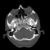
Anterior ethmoidal notch

Published
06 Oct 2019
41% complete
Annotated image
Case
Tracheal bronchus

Published
08 Oct 2019
92% complete
CT
Case
Normal elbow (AP acute flexion view)

Published
27 Jan 2020
43% complete
Diagram
Annotated image
Case
Illustration - displaced osteochondral lesion of the talar dome

Published
07 Feb 2020
22% complete
Diagram
Article
Congenital absence of the cruciate ligaments
Congenital absence of the cruciate ligaments is a rare condition, most of the knowledge being collated from case reports.
Epidemiology
Its prevalence is reported at 0.017 per 1000 live births 1,5.
Associations
The following features may also be present 2,3,5:
lateral femoral condyle hypopl...
Article
Pulvinar (disambiguation)
Pulvinar may refer to:
pulvinar thalamic nuclei (classically involved in variant Creutzfeldt-Jakob disease, see pulvinar sign)
Haversian fat pad of the hip (which covers the central non-articular part of the acetabulum)
Case
Vidian canal
Published
31 May 2020
48% complete
CT
Case
Rhomboid fossa on both clavicles

Published
06 Oct 2020
94% complete
X-ray
Article
International Frontal Sinus Anatomy Classification
International Frontal Sinus Anatomy Classification (IFAC) result from an expert consensus, developed to improve the ability of the surgeon to understand the possible variations of the frontal recess and frontal sinus anatomy.
Classification
anterior cells: push the drainage pathway of the fron...
Article
Supra agger frontal cell
Supra agger frontal cells are an anatomical variant of the paranasal sinuses, included in the International Frontal Sinus Anatomy Classification.
Gross anatomy
Anterior-lateral ethmoidal cell that extends into the frontal sinus. A small SAFC will only extend into the floor of the frontal sinus...
Article
Supra bulla cell
Supra bulla cells are an anatomical variant of the paranasal sinuses, included in the International Frontal Sinus Anatomy Classification.
Gross anatomy
Cell above the bulla ethmoidalis that does not enter the frontal sinus.
The supra agger cells push the drainage pathway anteriorly.


 Unable to process the form. Check for errors and try again.