
Items tagged “anatomy”
380 results found
Case
Mandibular canal
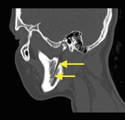
Published
19 Oct 2014
50% complete
CT
Annotated image
Case
Anatomy of the talus

Published
04 Nov 2014
41% complete
Diagram
Article
Talus
The talus (plural: tali 4), historically known as the astragalus, is a tarsal bone in the hindfoot that articulates with the tibia, fibula, calcaneus, and navicular bones. It has no muscular attachments and around 60% of its surface is covered by articular cartilage.
Gross anatomy
The talus h...
Article
Calcaneus
The calcaneus, also referred to as the calcaneum, (plural: calcanei or calcanea) is the largest tarsal bone and the major bone in the hindfoot. It articulates with the talus superiorly and the cuboid anteriorly and shares a joint space with the talonavicular joint, appropriately called the taloc...
Article
Patent foramen ovale
A patent foramen ovale (PFO) is a normal foetal interatrial connection which can persist into adult life. This can cause stroke due to paradoxical embolus.
Terminology
PFO is an anatomical variant due to persistence of a normal foetal structure rather than a malformation, in contradistinction ...
Article
Eustachian valve
The Eustachian valve, also known as the "valve of the inferior vena cava", is a ridge of variable thickness in the inferior right atrium. It is a remnant of a fetal structure that directed incoming oxygenated blood to the foramen ovale and away from the right atrium.
Radiographic features
In...
Article
Cervical spine ligaments
Cervical spine ligaments ordered from anterior to posterior include:
anterior longitudinal ligament (ALL)
anterior atlanto-occipital membrane
apical ligament
alar ligaments (paired)
cruciate ligament of the atlas
longitudinal band: joins the body of the axis to the foramen magnum
transver...
Article
Flexor retinaculum (wrist)
The flexor retinaculum (also known as the transverse carpal ligament) is a rectangular-shaped fibrous band located at the volar aspect of the hand, near the wrist.
Gross anatomy
The flexor retinaculum encloses and forms the roof of the carpal tunnel. The ulna aspect of the flexor retinaculum f...
Article
Arterial supply to the foot
Arterial supply to the foot can be divided into plantar and dorsal components.
Plantar arterial supply
Posterior tibial artery
gives off its calcaneal branch
then divides into the medial and lateral plantar arteries
Medial plantar artery
branch of the posterior tibial artery
smaller cal...
Article
Triquetrum
The triquetrum (also known as os triquetrum or - historically - as the triangular bone) is one of the carpal bones and forms part of the proximal carpal row.
Gross anatomy
Osteology
The triquetrum is wedge-shaped carpal bone located between the lunate and the pisiform. It has an oval facet fo...
Article
Radiological anatomy
Anatomy encompasses any part of an organism's structure, position, and interrelation. Radiopaedia.org aims to eventually cover the entire human anatomy, particularly in relation to the practice of radiology. All articles use standard anatomic conventional nomenclature and the anatomic position.
...
Case
Normal ileocecal junction

Published
27 Jan 2015
56% complete
Annotated image
CT
Fluoroscopy
Article
Anterior commissure
The anterior commissure (AC) is a transversely oriented commissural white matter tract that connects the two cerebral hemispheres along the midline. It is a very important anatomical landmark that connects different parts of the limbic system on both sides and plays a role in the interhemispheri...
Article
Agenesis of the right hepatic lobe
Agenesis of the right hepatic lobe is a rare variation in liver anatomy.
Radiographic features
absence of the right hepatic lobe
absence of right hepatic artery, right portal vein, and right hepatic biliary system
compensatory hypertrophy of the left hepatic lobe and caudate lobe
possible r...
Article
Agenesis of the left hepatic lobe
Agenesis of the left hepatic lobe is a rare variation in liver anatomy. It is clinically asymptomatic and discovered during imaging or surgery.
Radiographic features
absence of the left hepatic lobe (left of the falciform ligament, Couinaud segments 2 and 3)
absence of left hepatic artery, le...
Article
Oblique fissure
The oblique fissures (also called the major fissures or greater fissures) are bilateral structures in both lungs separating the lung lobes.
Gross anatomy
Right oblique fissure
The superior part of the right oblique fissure separates the right upper lobe from the right lower lobe and the infe...
Article
Ulna
The ulna (plural: ulnae) is one of the two long bones of the forearm, located medially in the supinated anatomic position. It has a larger proximal end and tapers to a smaller distal end (opposite to the radius).
Gross anatomy
Osteology
Prominent features of the ulna include:
proximal: olecr...
Article
Cloaca (urogenital)
The cloaca is the terminal portion of the hindgut. It is an embryonic structure (weeks 4-7) in which the distal ends of the gastrointestinal tract and urogenital system share a common channel. The most distal aspect of the cloaca is termed the cloacal membrane.
The cloaca, or portions of it, ca...
Article
Conus artery
The conus artery is a small early branch off the right coronary artery (RCA) circulation.
Gross anatomy
Supply
The artery has a variable distribution, but usually supplies a region of the anterior interventricular septum and the conus of the main pulmonary artery (hence its name).
Variant an...
Article
Lenticulostriate arteries
The lenticulostriate arteries, also known as anterolateral central arteries, are a collection of small perforating arteries arising from the anterior part of the circle of Willis and supplying the basal ganglia.
They are divided into:
medial lenticulostriate arteries
lateral lenticulostriate...


 Unable to process the form. Check for errors and try again.