
Items tagged “cerebellum”
37 results found
Case
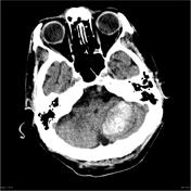
Hemangioblastoma - posterior fossa

Published
04 Feb 2011
64% complete
DSA (angiography)
CT
MRI
Article
Bilateral middle cerebellar peduncle lesions
Bilateral lesions of the middle cerebellar peduncles, resulting in the middle cerebellar peduncle sign, are uncommon and can be seen either in isolation (rare) or along with other regions of involvement.
Despite their relative rarity, they have a fairly long list of potential causes (see below)...
Article
Cerebellar hemorrhage
Cerebellar hemorrhages are a common form of intracerebral hemorrhage (ICH) and usually occur due to poorly controlled long-standing hypertension, although other causes also exist. When due to chronic hypertension, the stigmata of chronic hypertensive encephalopathy are often present (see: cerebr...
Case
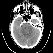
Cerebellar hemorrhage
Published
29 May 2012
92% complete
CT
Case
Medulloblastoma with obstructive hydrocephalus

Published
16 Apr 2013
77% complete
MRI
Case
Lhermitte-Duclos disease

Published
23 Apr 2013
68% complete
MRI
Case
Cerebellar hemorrhage

Published
10 Nov 2013
98% complete
CT
Case
Primary CNS lymphoma (cerebellar)

Published
22 Jan 2015
100% complete
CT
MRI
Article
Superior medullary velum
The superior (or anterior) medullary velum is a thin layer of tissue that is suspended between the superior cerebellar peduncles forming the roof of the fourth ventricle along with the inferior medullary velum. It is enclosed by pia mater dorsally and ependyma ventrally. The lingula of the vermi...
Article
Cerebellar tonsils
The cerebellar tonsils are ovoid structures on the inferomedial surface of each cerebellar hemisphere. They are attached to the underlying cerebellum by the tonsillar peduncle 1-4.
Gross Anatomy
Relations
medial: uvula of the vermis
superior: flocculonodular lobe
anterior: posterior surface...
Article
Middle cerebellar peduncle
The middle cerebellar peduncles, also known as the brachium pontis, are paired structures connecting the cerebellum to the pons.
Gross anatomy
The middle cerebellar peduncles contain afferent white matter projection fibers which originate in contralateral pontine nuclei. The corticopontocerebe...
Article
Inferior cerebellar peduncle
The inferior cerebellar peduncles are paired structures containing important white matter fiber tracts which connect the cerebellum to the medulla.
Gross anatomy
The inferior cerebellar peduncles are composed of a large restiform body and a small juxtarestiform body.
They contain the followi...
Article
Vermis
The vermis (pl: vermes) of the cerebellum is an unpaired medial structure that separates the cerebellar hemispheres. Its anatomy broadly follows that of the cerebellar hemispheres.
Gross anatomy
The vermis is separated into a small anterior lobe and a much larger posterior lobe by the primary...
Case
Cerebellar gangliocytoma

Published
11 Jan 2019
89% complete
MRI
Pathology
Case
Fogging phenomenon - cerebellar stroke

Published
29 Dec 2020
92% complete
CT
Case
Diffuse astrocytoma NOS in the cerebellum
Published
29 Jun 2022
95% complete
CT
MRI
Case
Acute cerebellitis

Published
11 Sep 2023
95% complete
MRI
Pathology


 Unable to process the form. Check for errors and try again.