Items tagged “diagram”
71 results found
Article
Ishikawa classification of venous involvement by pancreatic ductal adenocarcinoma
Ishikawa classification system describes the degree of involvement of adjacent portal vein and superior mesenteric vein by pancreatic ductal adenocarcinoma based on the caliber of the vein:
type I: normal
type II: smooth shift/displacement with normal caliber
type III: unilateral narrowing...
Case
Middle ear anatomy: diagrams

Published
03 Jun 2017
22% complete
Diagram
Case
Flexion teardrop fracture (illustration)

Published
25 Jul 2017
44% complete
Diagram
Case
Subdural hygroma illustration

Published
11 Sep 2017
29% complete
Diagram
Case
C1 posterior arch anomalies diagram: Currarino classification

Published
14 Sep 2017
19% complete
Diagram
Article
Transition zone (nerve)
The transition zone of a nerve, also known as the Obersteiner-Redlich zone, describes a region of a few millimeters where the myelin sheath changes from a central to peripheral type as enveloping glial cells are replaced by Schwann cells.
Gross anatomy
Transition zone locations 1:
CN V: 4 mm ...
Case
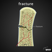
Fracture healing diagrams
Published
23 Dec 2017
30% complete
Diagram
Case
Sternocostal and interchondral joint anatomy (illustration)

Published
14 Feb 2019
22% complete
Diagram
Case
Menisci illustrations

Published
11 May 2019
20% complete
Diagram
Case
Twin to twin transfusion syndrome (TTTS) - illustration

Published
15 Jun 2019
29% complete
Diagram
Case
Twin reversed arterial perfusion - illustration

Published
21 Jun 2019
29% complete
Diagram
Article
Bunny waveform sign
Bunny waveform sign refers to the biphasic morphology of the pulsed wave Doppler spectral waveform in the vertebral artery in early (occult/latent or partial) subclavian steal phenomenon (sometimes called a "presteal" state, before it progresses to frank flow reversal). There is a sharp decelera...
Case
Castellvi classification - illustrations

Published
25 Sep 2019
16% complete
Diagram
Article
Lymph nodes
The lymph nodes (commonly shortened to nodes, and known as nodus lymphoideus in TA 4) collectively form one of the secondary lymphoid organs.
Gross anatomy
Macroscopically, a normal lymph node is a small ellipsoid structure, approximately 0.1 to 2.5 cm in maximal length 2,3. Nodes often posses...
Case
Femoroacetabular impingement (illustrations)

Published
19 Jan 2020
22% complete
Diagram
Case
Illustration - displaced osteochondral lesion of the talar dome

Published
07 Feb 2020
22% complete
Diagram
Article
Deep posterior tibiotalar ligament
The deep posterior tibiotalar ligament (DPTTL) is one of the two deep components and the strongest part of the deltoid ligament 1,2.
Gross anatomy
The deep posterior tibiotalar ligament is covered by the superficial posterior tibiotalar and tibiocalcaneal ligaments. It is a broad and thick lig...
Case
Bone lesion differential diagnosis - illustrations

Published
22 Jun 2020
35% complete
Diagram
Case
Jefferson fracture - illustration

Published
30 Sep 2020
19% complete
Diagram
Case
Orbital septum (diagram)

Published
31 Aug 2021
32% complete
Diagram