
Items tagged “hip”
162 results found
Case
Sickle cell disease

Published
22 Aug 2017
90% complete
MRI
X-ray
CT
Article
Iliopsoas bursitis
Iliopsoas bursitis usually presents as non-specific anterior hip pain and can be due to a number of causes, the three main causes being acute trauma, overuse injuries, and rheumatoid arthritis.
Pathology
Iliopsoas bursitis can occur primarily, e.g. overuse, secondary to a snapping iliopsoas te...
Case
Avascular necrosis - hip joint

Published
05 Feb 2019
75% complete
X-ray
Case
Enthesopathy vs enthesitis (diagram)
Published
09 Jul 2019
44% complete
Diagram
Case
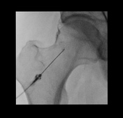
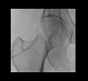
Hip arthrogram injection (fluoroscopic guided)
Published
05 Jan 2020
65% complete
Fluoroscopy
Case
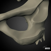
Femoroacetabular impingement (illustrations)

Published
19 Jan 2020
22% complete
Diagram
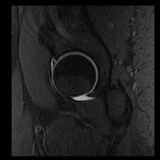
Case
Ischial apophyseal stress injury

Published
15 Mar 2020
92% complete
MRI
Case
Hip arthrogram injection (fluoroscopic guided)

Published
01 Apr 2020
66% complete
Fluoroscopy
Case
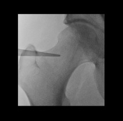
Hip anesthetic arthrogram injection (fluoroscopic guided)
Published
26 Apr 2020
66% complete
Fluoroscopy
Case
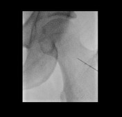
Hip anesthetic arthrogram injection (fluoroscopic guided)
Published
02 May 2020
65% complete
Fluoroscopy
Case
Hip anesthetic arthrogram injection (fluoroscopic guided)
Published
29 Apr 2020
66% complete
Fluoroscopy
Case
Focal hip chondrolabral separation
Published
26 Apr 2020
92% complete
MRI
Article
Hip joint injection (technique)
Hip joint injections can be performed with a variety of image guidance, including fluoroscopy and ultrasound, which are used to administer MRI arthrogram injectate, or a steroid containing anesthetic arthrogram injectate.
Indications
MRI: labral injury
anesthetic
pain/arthropath...
Article
Hip microinstability
Hip microinstability is a multifactorial disorder referred to as painful, excessive mobility of the femoral head within the acetabulum 1,2. It results from impaired joint stability, secondary to functional and architectural abnormalities 1,2. AB-HEER test on clinical examination remains the mos...
Article
Ilioischial line
The ilioischial line, also known as the Köhler line, is a radiologic feature seen in the AP pelvis view and serves to assess the posterior acetabular column 1-4.
Measurement
The ilioischial line takes its course along the lateral border of the obturator foramen to the medial border of the ilia...
Article
Carpet lesion
Carpet lesion is a term for focal chondral delamination, where articular cartilage is peeled off the subchondral bone plate as a result of shearing forces. It is a frequent finding on hip arthroscopy and is associated with femoroacetabular impingement 1,2, particularly cam morphology 6.
Termino...
Article
Cam morphology (femoroacetabular impingement)
Cam morphology refers to an abnormal morphology of the femoral head-neck junction interlinked with an osseous asphericity of the femoral head. It is one possible cause of femoroacetabular impingement (FAI).
Terminology
Cam morphology is also commonly referred to as 'cam deformity', 'cam lesion...
Article
Pincer morphology (femoroacetabular impingement)
Pincer morphology refers to an abnormality of the acetabulum, in particular, acetabular overcoverage, which can be focal or global and is one cause of femoroacetabular impingement.
Terminology
Pincer morphology is also referred to as 'pincer deformity', though according to the Warwick agreeme...
Article
Ischial spine sign
The ischial spine sign is a radiographic sign on the AP view of the pelvis, for the diagnosis of acetabular retroversion, which poses a form of pincer morphology and predisposes to femoroacetabular impingement.
Acetabular retroversion seems to be a result of a rotation of the entire acetabular ...
Article
ARCO classification of femoral head osteonecrosis
The ARCO classification (Association Research Circulation Osseous classification) is one of the staging systems used to assess femoral head osteonecrosis. It was created in 1994 and periodically revised. The most recent revision from 2019 2 includes using radiographs and MRIs.
Classification
s...


 Unable to process the form. Check for errors and try again.