Items tagged “illustration”
68 results found
Case
Glenoid - Gray's anatomy illustration

Published
27 Feb 2010
41% complete
Diagram
Article
Psoas major muscle
The psoas major muscle (usually shortened to just the psoas muscle) is one of the muscles of the posterior abdominal wall and lies not in the retroperitoneum but posterior to it, in the iliopsoas compartment.
Summary
origin: vertebral bodies, intervertebral discs and transverse processes of T1...
Case
Ductus venosus flow waveform

Published
14 May 2011
47% complete
Diagram
Case
Radioulnar synostosis - illustration of functional difficulties

Published
24 Oct 2012
29% complete
Diagram
Case
Normal epiglottis location (illustration)

Published
02 Feb 2013
29% complete
Annotated image
Case
Danger space potential location (illustration)

Published
02 Feb 2013
44% complete
Diagram
Article
Medical illustrations and diagrams
High-quality medical illustrations and diagrams form an important part of Radiopaedia.org.
Attribution
All illustrations should have appropriate attribution in the case findings.
We strongly favor original illustrations. This makes Radiopaedia unique and contributes to what is available for...
Article
Flexor digitorum superficialis muscle
Flexor digitorum superficialis (FDS) muscle, also known as flexor digitorum sublimis muscle, is a muscle in the second (intermediate) layer of the anterior compartment of the forearm. It splits into four tendons, passes through the carpal tunnel under the flexor retinaculum. At the level of the ...
Article
Flexor digitorum profundus muscle
The flexor digitorum profundus (FDP) muscle makes up the third layer of the anterior compartment of the forearm along with the flexor pollicis longus muscle. It passes through the carpal tunnel and is one of the extrinsic muscles of the hand. The flexor digitorum profundus muscle is considered a...
Article
Flexor pollicis longus muscle
The flexor pollicis longus (FPL) muscle is one of the two muscles that make up the third layer of the anterior compartment of the forearm, along with the flexor digitorum profundus muscle. It is a deep muscle under the abductor pollicis brevis muscle. It passes through the carpal tunnel. It is o...
Case
Triangular fibrocartilage complex (TFCC) anatomy illustrations

Published
08 Aug 2014
32% complete
Diagram
Case
Anatomy of the talus

Published
04 Nov 2014
41% complete
Diagram
Case
Weber classification of ankle fractures

Published
14 Apr 2015
44% complete
Diagram
Case
Scapula: anatomy diagrams

Published
19 May 2015
19% complete
Diagram
Case
Supra-acetabular fossa and notch (diagrams)

Published
19 May 2015
32% complete
Diagram
Case
Modic-type endplate changes: diagram

Published
19 May 2015
29% complete
Diagram
Case
Tibialis posterior tendon anatomy: diagram

Published
19 May 2015
19% complete
Diagram
Case
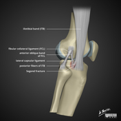
Segond fracture (diagram)
Published
12 Jun 2015
41% complete
Diagram
Case
Brain ventricle anatomy diagram
Published
23 Jun 2015
29% complete
Diagram
Case
Renal trauma grading diagrams

Published
23 Jun 2015
35% complete
Diagram