
Items tagged “normal variant”
116 results found
Article
Os hamuli proprium
The hook of the hamate has its own ossification center, which may fail to fuse with the body of the hamate, creating an ossicle. This ossicle is referred to as an os hamuli proprium or unfused hamulus
In some instances, this can be difficult to distinguish from a fracture of the hook, but is s...
Article
Accessory ossicles of the wrist (mnemonic)
The accessory ossicles of the wrist can be easily recalled with the mnemonics:
LOTTEO 1
HOTELS
Mnemonics
LOTTEO
L: lunula
O: os styloideum (carpal boss)
T: (os) triangulare
T: (os) trapezium secondarium
E: (os) epilunate
O: os hamuli proprium
HOTELS
H: (os) hamuli proprium
O: os tri...
Case
Accessory navicular bone

Published
07 Jul 2013
83% complete
MRI
Article
Accessory left atrial appendage
An accessory left atrial appendage is a frequent fortuitous finding in cardiac imaging, encountered in ~10% of patients. They are more often seen as a small diverticular structure projecting from the right upper side of the left atrial wall.
Differential diagnosis
it must not be confused with ...
Case
Anterior cerberal artery fenestration

Published
26 Jul 2013
100% complete
MRI
Case
Bovine arch

Published
29 Jul 2013
71% complete
CT
Case
Persistent left superior vena cava

Published
30 Jul 2013
77% complete
CT
X-ray
Case
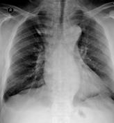
Azygos lobe
Published
05 Aug 2013
85% complete
X-ray
Case
Bilateral sternalis muscle at mammography

Published
10 Aug 2013
68% complete
Mammography
Case
Cleft epiphysis - tibia

Published
11 Aug 2013
88% complete
X-ray
Case
Cleft ulnar epiphysis and persistent radial styloid

Published
11 Aug 2013
75% complete
X-ray
Case
Bicornuate uterus

Published
04 Sep 2013
74% complete
MRI
Article
Ramus intermedius artery
The ramus intermedius is a variant coronary artery resulting from trifurcation of the left main coronary artery 1. It is present in ~20% (range 15-30%) 2,3 of the population.
It can have a course similar to the obtuse marginal branches of the left circumflex artery or the diagonal branches of t...
Case
Fetal origin of the posterior cerebral artery

Published
21 Jan 2014
59% complete
CT
Case
Chilaiditi sign

Published
23 Jan 2014
91% complete
X-ray
Case
Dense calcaneal apophysis (normal)

Published
22 Apr 2014
82% complete
X-ray
Case
Chilaiditi sign on CT

Published
25 Feb 2014
80% complete
CT
Case
Coracoclavicular joint - bilateral

Published
17 Apr 2014
80% complete
X-ray
Annotated image
Case
Accessory occipital-C1 articulation

Published
09 Jun 2014
74% complete
CT
Case
Meniscal ossicle

Published
08 Aug 2014
92% complete
X-ray
MRI


 Unable to process the form. Check for errors and try again.