
Items tagged “normal variant”
116 results found
Case
Subclavius posticus muscle - an accessory muscle

Published
09 Sep 2014
84% complete
MRI
Annotated image
Case
Tug lesion

Published
14 Sep 2014
85% complete
X-ray
Case
Osteochondritis dissecans of the elbow and synovial osteochondromatosis

Published
26 Oct 2014
79% complete
Annotated image
X-ray
Case
Isolated spade phalanx

Published
16 Mar 2015
94% complete
X-ray
Case
Left superior intercostal vein - aortic nipple

Published
25 Mar 2015
83% complete
Annotated image
X-ray
Case
Occipital spur
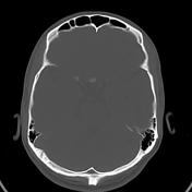
Published
06 May 2015
77% complete
CT
Case
Supra-acetabular fossa and notch (diagrams)

Published
19 May 2015
32% complete
Diagram
Case
Fetal rhombencephalon

Published
12 Jun 2015
63% complete
Ultrasound
Annotated image
Article
Areae gastricae
Areae gastricae are a normal finding on double contrast images of the stomach.
Radiographic features
fine reticular network of barium-coated grooves between 1-5 mm islands/areas of gastric mucosa
may be seen in ~70-80% of patients if there is adequate high-density barium coating of the stomac...
Article
Glycogenic acanthosis
Glycogenic acanthosis is a benign finding on esophagography in elderly patients.
Epidemiology
It most commonly occurs in patients >40 years of age and incidence and numbers of lesions increase by age. No gender predilection exists. Typically patients are asymptomatic.
Pathology
It occurs fr...
Case
Azygos lobe

Published
21 Oct 2015
65% complete
CT
X-ray
Case
Cavum velum interpositum

Published
17 Oct 2015
80% complete
MRI
Case
Anteromedial meniscofemoral ligament (of the medial meniscus)

Published
06 Nov 2015
89% complete
MRI
Case
Compound calyx (normal variant)

Published
25 Nov 2015
91% complete
Fluoroscopy
Annotated image
Article
Cumulus oophorus
Cumulus oophorus refers to an appearance in the ovary in which multiple granulosa cells enlarge around a developing oocyte. These support cells ("cumulus cells") serve multiple functions in the maturation of the oocyte. They may occasionally be seen during a pelvic ultrasound, and should not be ...
Article
Gastrointestinal nodular lymphoid hyperplasia
Gastrointestinal nodular lymphoid hyperplasia is a type of nodular lymphoid hyperplasia that can be found elsewhere in the body. It is formed out of gut-associated lymphoid tissue (GALT), and most often is a diagnostic dilemma for radiologists in the stomach and terminal ileum.
Pathology
Gut-a...
Case
Gastrointestinal nodular lymphoid hyperplasia
Published
18 Apr 2016
75% complete
Barium
Annotated image
Case
Carpal boss

Published
16 Jun 2016
66% complete
X-ray
Case
Right sided aortic arch with mirror image branching and absent celiac trunk

Published
15 Jul 2016
95% complete
CT
Case
Os naviculare: diagrams

Published
23 Aug 2016
25% complete
Diagram


 Unable to process the form. Check for errors and try again.