
Items tagged “pelvis”
126 results found
Article
Tombstone iliac wings
Tombstone iliac wings, also referred to as Mickey Mouse ears pelvis, is an imaging descriptor for the iliac wings of individuals with achondroplasia 1. These are seen to be small and squared and have been likened to the appearance of tombstones or the ears of Mickey Mouse.
Case
Myxoid chondrosarcoma

Published
12 Feb 2010
62% complete
X-ray
MRI
CT
Case
Osteochondroma

Published
24 Feb 2010
75% complete
X-ray
CT
MRI
Case
Ankylosing spondylitis

Published
20 Oct 2010
60% complete
X-ray
Case
Avulsion fracture - anterior superior iliac spine

Published
20 Oct 2010
60% complete
X-ray
Annotated image
Case
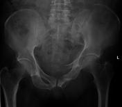
Pubic symphysis widening

Published
20 Oct 2010
66% complete
X-ray
Case
Infectious myositis

Published
20 Oct 2010
62% complete
MRI
Case
Osteitis pubis

Published
20 Oct 2010
63% complete
X-ray
Case
Perirectal fistula and abscess

Published
20 Oct 2010
59% complete
CT
Annotated image
Case
Radiation osteonecrosis

Published
20 Oct 2010
22% complete
X-ray
Case
Malgaigne fracture
Published
23 Oct 2010
68% complete
X-ray
CT
Article
Urinary bladder hernia
Herniation of the urinary bladder is a relatively uncommon but not a rare condition. It occurs when the urinary bladder or ureter herniates into the inguinal canal, scrotal sac or femoral canal. Herniations through ischiorectal, obturator, or abdominal wall openings have also been described. Bla...
Article
Peritoneal inclusion cyst
Peritoneal inclusion cysts, also known as peritoneal pseudocysts, are a type of cyst-like structure that appears in relation to the peritoneal surfaces and results from a non-neoplastic reactive mesothelial proliferation.
Terminology
The nomenclature for this condition can be confusing due to ...
Article
Presacral space
The presacral space is located between the rectum and the sacrococcygeal part of the spine.
Gross anatomy
Contents
The presacral space contains a variety of tissue:
fat
mesenchymal tissue
lymph nodes
nerve plexuses
blood vessels
Boundaries
superior - peritoneal reflections
inferior - ...
Case
Lupus osteonecrosis

Published
18 Apr 2011
38% complete
X-ray
Article
Parturition-induced pelvic instability
Parturition-induced pelvic instability is a rare condition seen in women following vaginal delivery.
Epidemiology
The incidence of symphyseal rupture after vaginal delivery ranges from one in 600 to one in 30,000 deliveries 1.
Predisposing factors include multiparity, complicated delivery, ...
Article
Common iliac artery
The common iliac arteries (CIA) are the large paired terminal branches of the abdominal aorta.
Gross anatomy
Origin
The abdominal aorta bifurcates anterolateral (to the left side) of the L4 vertebra into the right and left common iliac arteries.
Course
The common iliac arteries (CIAs) ente...
Article
Internal iliac artery
The internal iliac artery (also known as the hypogastric artery, but internal iliac is the accepted term in the TA) is the smaller terminal branch of the common iliac artery. It supplies the pelvic walls, pelvic viscera, external genitalia, perineum, buttock and medial part of the thigh.
Gross...
Case
Pelvic lipomatosis

Published
10 Feb 2012
59% complete
CT
Article
Epididymis
The epididymis (plural: epididymides) is situated adjacent to the testis within the scrotal sac. Its primary function is the collection, maturation and transport of sperm via the ductus deferens.
Gross anatomy
The epididymis is an elongated structure, posterolateral to the testis. It can be su...


 Unable to process the form. Check for errors and try again.