Items tagged “pocus”
122 results found
Article
Ultrasound of the knee
Ultrasound of the knee allows high-resolution imaging of superficial knee anatomy while simultaneously allowing dynamic evaluation of some of the tendons and ligaments. Knee ultrasound is somewhat limited compared with ultrasound examinations of other joints because the cruciate ligaments and th...
Article
Cardiovascular shunts
Cardiovascular (cardiac) shunts are abnormal connections between the pulmonary and systemic circulations. Most commonly they are the result of congenital heart disease.
Pathology
Blood can either be shunted from the systemic circulation to pulmonary circulation (i.e. 'left-to-right shunt') or ...
Article
Pneumothorax (ultrasound)
Pneumothorax is a serious potential consequence of blunt thoracic trauma and, if misdiagnosed, it may quickly become life-threatening.
For a discussion on epidemiology, clinical presentation, pathology, and treatment and prognosis please see the main pneumothorax article.
Radiographic feature...
Article
Asteroid hyalosis
Asteroid hyalosis is a degenerative condition of the eye where there is an accumulation of calcium soaps in the vitreous chamber.
Epidemiology
The prevalence increases with age from 0.2% in 43-54 year-olds to 2.9% in 75-86 year-olds. The overall prevalence is 1.2%. It is commonly unilateral an...
Article
Choroidal detachment
Choroidal detachment is a detachment of the choroid from the underlying sclera due to the accumulation of fluid in the suprachoroidal space generally due to increased intraocular pressure (IOP), as observed in some settings:
choroidal effusion
transudative: trauma
exudative: fluid accumulatin...
Article
Abdominal paracentesis
An abdominal paracentesis (plural: paracenteses), more commonly referred to as an ascitic tap, is a procedure that can be performed to collect peritoneal fluid for analysis or as a therapeutic intervention.
Indications
diagnostic: especially for newly-diagnosed ascites
determine etiology of a...
Article
Rapid ultrasound in shock
The rapid ultrasound in shock (RUSH) protocol is a structured point-of-care ultrasound (POCUS) examination performed at the time of presentation of a shocked patient. It is a more detailed and longer exam than the FAST scan, with the aim to differentiate between hypovolemic, cardiogenic, obstruc...
Article
Sonographic approach to dyspnea (mnemonic)
This mnemonic will help with the sonographic approach to the critically ill patient with dyspnea:
CHEST
Mnemonic
C: collapsed lung (pneumothorax)
absence of anterior lung sliding, lung pulse, B-lines, or z-lines
these artifacts arise from the pleural interface; their presence would rule ou...
Article
Ultrasound-guided peripheral intravenous cannulation
Peripheral intravenous cannulation under ultrasound guidance is the placement of a cannula into a peripherally-located vein under the direct vision of ultrasound. This process allows the cannulation of veins that are unable to be visualized or palpated without ultrasound. In trained individuals ...
Article
Vitreous hemorrhage
Vitreous hemorrhage refers to bleeding into the vitreous humor.
Epidemiology
Vitreous hemorrhage has an incidence of approximately 7 in 100,000 1,2.
Clinical presentation
The most common clinical presentation is with sudden, painless visual loss to varying degrees of severity 2. Associated...
Article
Normal hepatic vein Doppler
The hepatic veins have a characteristic spectral Doppler waveform. Alterations in the normal hepatic vein waveform may reveal or confirm abnormalities in the heart or liver.
Terminology
The shape of the hepatic vein spectral Doppler waveform is primarily determined by pressure changes in the r...
Article
Aortic valve regurgitation
Aortic valve regurgitation, also known as aortic valve insufficiency or aortic valve incompetence, is a valvulopathy that describes leaking of the aortic valve during diastole that causes blood to flow in the reverse direction from the aorta and into the left ventricle.
Epidemiology
Aortic reg...
Article
Aorto-ventricular tunnel
Aorto-ventricular tunnel (AVT) is an extremely rare form of congenital heart disease, representing an anomalous extracardiac communication between the ascending aorta and the left or right ventricles.
Terminology
In most cases the anomalous communication is between the aorta and the left ventr...
Case
Normal anterior lung (ultrasound)
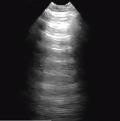
Published
30 Jun 2018
54% complete
Ultrasound
Case
Inferior vena cava (ultrasound)
Published
02 Jul 2018
60% complete
Ultrasound
Article
Bedside lung ultrasound in emergency (approach)
Bedside lung ultrasound in emergency (BLUE) is a basic point-of-care ultrasound (POCUS) examination performed for undifferentiated respiratory failure at the bedside, immediately after the physical examination, and before echocardiography.
The protocol is simple and dichotomous, and takes fewer...
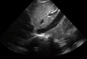
Case
Pneumonia - ultrasound

Published
25 Aug 2018
66% complete
Ultrasound
Case
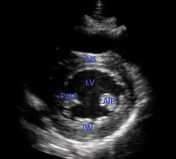
Parasternal long axis view - normal (transthoracic echocardiography)

Published
25 Aug 2018
54% complete
Ultrasound
Case
Parasternal short axis view - normal (transthoracic echocardiography)
Published
25 Aug 2018
54% complete
Ultrasound
Case
Cardiogenic pulmonary edema (ultrasound)

Published
29 Aug 2018
75% complete
Ultrasound