
Items tagged “portal hypertension”
16 results found
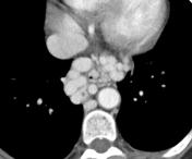
Case
Gastroesophageal varices

Published
14 Dec 2010
92% complete
CT
Case
Esophageal varices
Published
18 Nov 2011
42% complete
CT
Case
Splenorenal collaterals in portal hypertension

Published
28 Nov 2011
80% complete
CT
Case
Portal vein thrombosis

Published
23 Apr 2012
80% complete
CT
Case
Esophageal duplication cyst

Published
01 May 2013
65% complete
CT
Article
Idiopathic non-cirrhotic portal hypertension
Idiopathic non-cirrhotic portal hypertension is the clinical diagnosis of exclusion featuring portal hypertension without hepatic cirrhosis, vascular obstruction, schistosomiasis, or a variety of other chronic liver diseases.
Terminology
Prior terms for this entity include non-cirrhotic portal...
Case
Noncirrhotic idiopathic portal hypertension

Published
08 Mar 2015
68% complete
CT
Ultrasound
Article
TIPS evaluation
TIPS evaluation is useful to ensure that a transjugular intrahepatic portosystemic shunt (TIPS) is working properly and that no stenosis has occurred within the stent. Ultrasound is often used as a first-line modality.
Radiographic features
Ultrasound
The normal TIPS should show color Doppler...
Case
Gamna-Gandy bodies (nodules)

Published
09 Mar 2016
92% complete
MRI
Case
Gamna Gandy bodies
Published
17 Dec 2016
63% complete
Ultrasound
Case
Hepatopulmonary syndrome

Published
18 Jun 2017
71% complete
CT
Case
Pancreatic pseudocyst with splenic vein compression

Published
06 Mar 2018
98% complete
CT
Article
Gastric varix
Gastric varices are an important portosystemic collateral pathway, occurring in ~20% of patients with portal hypertension. They are considered distinct from esophageal varices in that they have a propensity to hemorrhage at comparatively lower portal pressures 1, and are also associated with hig...
Article
Denver shunt
A Denver shunt, or peritoneovenous shunt, is a device used to shunt ascites to the superior vena cava in patients with refractory ascites.
The proximal end is located in the peritoneal cavity and the distal end in the superior vena cava, with a subcutaneous course in the anterior chest wall. It...
Article
Serum ascites albumin gradient
The serum–ascites albumin gradient (SAAG) is the difference between the concurrently obtained serum albumin concentration and the albumin concentration of the ascitic fluid obtained during paracentesis.
Pathology
A difference ≥1.1 grams/deciliter (g/dL) indicates portal hypertension as the li...
Case
Pancreatic pseudocyst

Published
03 Oct 2022
80% complete
MRI


 Unable to process the form. Check for errors and try again.