
Items tagged “ref”
30 results found
Article
Pelvic peritoneal space
The pelvic peritoneal space is the inferior reflection of the peritoneum over the fundus of the urinary bladder and the front of the rectum at the junction of its middle and lower thirds. In females, the reflection is also over the anterior and posterior surface of the uterus and the upper poste...
Article
Traumatic aortic injury in the exam
Getting a film with traumatic aortic injury in the exam is one of the many exam set-pieces that can be prepared for.
This is one of the cases you should look and not speak for 10 seconds as there tends to be a lot of findings on the film of patients with a traumatic aortic injury.
Description...
Article
Diastolic pseudogating
Diastolic pseudogating appears as periodic bright and dark signal in arteries such as the aorta as one progresses through a series of images. Synchronisation of the cardiac cycle and the pulse sequence results in high signal in the artery during diastole when blood is relatively stationary and l...
Case
Gastric mucinous adenocarcinoma

Published
17 Jun 2013
71% complete
CT
Case
Carcinoma of the cervix
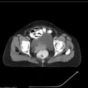
Published
21 Jun 2013
62% complete
CT
Article
Retrotracheal space
The retrotracheal space (or Raider triangle) is a radiolucent mediastinal space best seen on lateral chest x-rays. It is normally triangular in shape but can vary greatly in size and shape depending on the patient's body habitus and lung volume 1.
Boundaries
anterior: posterior tracheal stripe...
Article
Onion bulb formation
Onion bulb formation is a histopathologic finding in hypertrophic neuropathies such as Charcot-Marie-Tooth type 1A (CMT1A) and chronic inflammatory demyelinating polyneuropathy (CIDP).
Pathology
Onion bulb formations are concentric layers of Schwann cell processes and collagen surrounding axon...
Case
Aberrant right subclavian artery

Published
13 Aug 2016
98% complete
CT
X-ray
Article
Raghib syndrome
Raghib syndrome is a rare developmental complex. It consists of:
persistence of the left superior vena cava
coronary sinus ostial atresia
atrial septal defect
It has also been associated with other congenital malformations including ventricular septal defects, enlargement of the tricuspid an...
Article
Superior vena cava obstruction (grading)
SVC obstruction can cause SVC syndrome which is the most common condition affecting this vessel. It can be secondary to extrinsic compression or intraluminal thrombosis/stenosis. Collateral pathways, with the azygos vein being the most important collateral vessel, form in response to severe narr...
Article
Angiomyofibroblastoma-like tumor of scrotum
Angiomyofibroblastoma-like tumor of the scrotum is a rare, well-defined, slow growing mesenchymal extratesticular nonepididymal tumor rarely seen in the perineum or scrotum of older male patients. A similar tumor can occur in females in the vulval region.
Epidemiology
In males, they are seen ...
Article
Obstruction of nasolacrimal drainage apparatus
Obstruction of nasolacrimal drainage apparatus results in epiphora and can be primary or secondary, congenital or acquired. Obstruction can occur at canalicular, lacrimal saccular, or nasolacrimal ductal (post-saccular) levels.
Causes of obstruction
Congenital obstruction
persistence of the m...
Article
Anterior lacrimal crest
The anterior lacrimal crest is a bony projection on the frontal process of the maxilla continuous with the orbital rim which creates the lateral margin of the lacrimal sac fossa. The medial palpebral ligament is attached to anterior lacrimal crest.
Immediately anterior to the anterior lacrimal ...
Article
Posterior lacrimal crest
The posterior lacrimal crest is a bony projection on the lacrimal bone which creates the medial margin of the lacrimal sac fossa.
See also
anterior lacrimal crest
Article
Lacrimal sac fossa
The lacrimal sac fossa is an excavated fossa in the inferior aspect of the anteromedial orbital wall which contains the lacrimal sac. It is bounded by the anterior and posterior lacrimal crests of the maxillary and lacrimal bones, respectively. In adults, it measures approximately 8-9 mm anterop...
Article
Maxillary line
The maxillary line is a mucosal projection along the lateral nasal wall corresponding to lacrimomaxillary suture externally. The midportion of the line is called "M point". During endoscopic sinus and orbital procedures the maxillary line and M-point are very important and useful landmarks in pa...
Article
Physis
The physes (singular: physis) or growth plates are found in bones that undergo endochondral ossification.
Radiographic features
The physis appears as a radiolucent line in skeletally-immature patients located between the metaphysis and epiphysis. It contains zones of mesenchymal cells in va...
Article
Double contour cartilage line
The double contour cartilage line is a sonographic sign specific for gout, which is characterized by an echogenic line on the outer surface of the joint cartilage parallel to the subchondral bone secondary to deposition of monosodium urate crystals on the surface of hyaline articular cartilage.
Article
Torus tubarius
Torus tubarius (plural: tori tubarii) or cushion of the auditory canal is a mucosal elevation in the lateral aspect of the nasopharynx, formed by the underlying pharyngeal end of the cartilaginous portion of the Eustachian tube. The opening of the Eustachian tube is anterior to the torus tobariu...
Article
Connolly procedure
The Connolly procedure is performed by an open posterior approach and involves transferring the infraspinatus with a portion of greater tuberosity into the defect, rendering the defect extra-articular; although this procedure restores the stability, it reduces the shoulder range of movement. The...


 Unable to process the form. Check for errors and try again.