Items tagged “spleen”
95 results found
Article
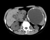
Splenic cyst
Splenic epithelial cysts, also known as splenic epidermoid cysts or primary splenic cysts, are unilocular fluid lesions with thin and smooth walls and no enhancement. They represent ~20% of cysts found in the spleen and are usually an innocuous incidental imaging finding.
Note that most (~80%) ...
Case
Hydatid of the spleen

Published
26 May 2008
58% complete
X-ray
CT
Case
Gaucher disease - massive splenomegaly

Published
05 Apr 2009
50% complete
MRI
Article
Increased splenic density
Increased splenic density can be due to a number of processes. The density may be due to calcification (most common) or other compounds (iron, Thorotrast), and can be seen (often incidentally) on abdominal radiographs and CT. On CT the usual splenic attenuation is 35-55 HU or ~10 HU 6 lower than...
Case
Splenunculus

Published
14 Aug 2009
65% complete
CT
Article
Splenunculus
Splenunculi, also known as supernumerary spleens, accessory spleens, or splenules, are small nodules of spleen that are separate from the rest of the organ.
Epidemiology
They are common, seen in up to 16% of CTs of the abdomen and up to 30% of autopsies 2.
Pathology
Accessory spleens are c...
Article
Splenosis
Splenosis is one type of ectopic splenic tissue (the other being accessory spleen). It is an acquired condition and is defined as autoimplantation of one or more focal deposits of splenic tissue in various compartments of the body.
Pathology
Etiology
Abdominal splenosis is seen after abdomina...
Case
SMA thromboembolism and splenic infarcts

Published
03 Sep 2009
74% complete
CT
Case
Inguinal node metastasis and wandering spleen

Published
14 Sep 2009
92% complete
CT
Case
Sickle cell disease

Published
24 Nov 2009
53% complete
CT
Article
Autosplenectomy
Autosplenectomy denotes spontaneous infarction of the spleen with resulting hyposplenism.
Epidemiology
Autosplenectomy is most frequently encountered in patients with homozygous sickle cell disease, although it has also been reported in pneumococcal septicemia 1, and systemic lupus erythematos...
Article
Splenic lesions and anomalies
There are a number of splenic lesions and anomalies:
Gamuts
hypervascular splenic lesions
Congenital anomalies
accessory spleen
wandering spleen
asplenia
polysplenia
bipartite spleen
splenogonadal fusion
retrorenal spleen
Mass lesions
Benign mass lesions
splenic cyst
splenic pseudo...
Case
Miliary tuberculosis (pathology)

Published
18 Feb 2010
68% complete
Pathology
Case
Spleen - normal arterial enhancement

Published
05 Apr 2010
53% complete
CT
Case
Seurat spleen - blunt splenic trauma

Published
10 Apr 2010
91% complete
DSA (angiography)
Annotated image
Article
Seurat spleen
Seurat spleen is an angiographic appearance seen following blunt trauma to the spleen. Multiple small punctate regions of intraparenchymal contrast extravasation lead to a spotted appearance.
Pathology
Several mechanisms are thought to to attribute to this appearance which include sinusoidal s...
Case
Splenic arterial aneurysm

Published
01 Jun 2010
63% complete
Ultrasound
Case
Splenic hemangioma

Published
25 Jun 2010
67% complete
Ultrasound
CT
Case
Sickle cell disease

Published
05 Aug 2010
85% complete
Ultrasound
Case
Hydatid cyst of the spleen
Published
28 Aug 2010
62% complete
CT