
Items tagged “trauma”
698 results found
Case
4th metacarpal fracture

Published
10 Oct 2009
88% complete
X-ray
Case
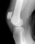
Lipohaemarthrosis

Published
05 Nov 2009
72% complete
X-ray
Case
Lipohaemarthrosis - shoulder

Published
05 Nov 2009
90% complete
Annotated image
X-ray
Case
Patellar tendon rupture
Published
05 Nov 2009
75% complete
X-ray
Article
Morel-Lavallée lesion
Morel-Lavallée lesions are closed degloving injuries associated with severe trauma which then present as haemolymphatic collections or masses. MRI and ultrasound are useful modalities for evaluation.
Terminology
The lesions classically occur over the greater trochanter of the femur 1. Morel-La...
Case
Biloma

Published
02 Dec 2009
62% complete
CT
Article
Maisonneuve fracture
Maisonneuve fracture refers to a combination of a fracture of the proximal fibula together with an unstable ankle injury (widening of the ankle mortise on x-ray), often comprising ligamentous injury (distal tibiofibular syndesmosis, deltoid ligament) and/or fracture of the medial malleolus. It i...
Case
Distal phalanx fracture

Published
01 Mar 2010
60% complete
X-ray
Article
Segond fracture
Segond fracture is an avulsion fracture of the knee that involves the lateral aspect of the tibial plateau and is very frequently (~75% of cases) associated with disruption of the anterior cruciate ligament (ACL). On the frontal knee radiograph, it may be referred to as the lateral capsular sign...
Article
Seurat spleen
Seurat spleen is an angiographic appearance seen following blunt trauma to the spleen. Multiple small punctate regions of intraparenchymal contrast extravasation lead to a spotted appearance.
Pathology
Several mechanisms are thought to to attribute to this appearance which include sinusoidal s...
Case
Jefferson fracture

Published
03 May 2010
98% complete
CT
X-ray
Annotated image
Case
Proximal interphalangeal joint dislocation - fifth finger

Published
03 May 2010
75% complete
X-ray
Case
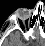
Vitreous haemorrhage - traumatic
Published
12 May 2010
59% complete
CT
Article
CT hypoperfusion complex
CT hypoperfusion complex refers to the predominantly abdominal imaging features that occur in the context of profound hypotension. Multiple abdominal organs can display atypical appearances not related to the initial trauma but reflect alterations in perfusion secondary to hypovolaemia which aff...
Case
Urinary bladder rupture

Published
24 May 2010
82% complete
Ultrasound
Case
Multitrauma with thoracic and intracranial injuries

Published
28 May 2010
80% complete
CT
Case
Transorbital penetrating brain injury

Published
29 May 2010
81% complete
CT
MRI
Article
Perilunate dislocation
Perilunate dislocations and perilunate fracture-dislocations are potentially devastating closed wrist injuries that are often missed on initial imaging.
These injuries involve dislocation of the carpus relative to the lunate which remains in normal alignment with the distal radius. They should...
Article
Lunate dislocation
Lunate dislocations are an uncommon traumatic wrist injury that require prompt management and surgical repair. The lunate is displaced and rotated volarly. The rest of the carpal bones are in a normal anatomic position in relation to the radius.
These should not be confused with perilunate disl...
Article
Scapholunate dissociation
Scapholunate dissociation, also known as rotary subluxation of the scaphoid, refers to an abnormal orientation of the scaphoid relative to the lunate and implies severe injury to the scapholunate interosseous ligament and other stabilising ligaments.
Carpal dissociation implies carpal instabili...


 Unable to process the form. Check for errors and try again.