
Items tagged “ultrasound”
506 results found
Article
Doppler angle correction
Doppler angle correction refers to an imaging post-processing method used to adjust for the effects of insonation angle on the Doppler shift.
Measurement of flow velocity with Doppler imaging is dependent on the angle between the ultrasound beam and the target (insonation angle), with the maxim...
Article
Wall filter
The wall filter in ultrasound is a way of filtering out low or high frequency Doppler signals. In clinical ultrasound, it is usually used to filter out very low frequencies that may add noise to a spectral Doppler waveform.
A typical use is removing the low frequency reverberation of an arteria...
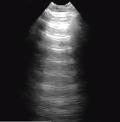
Case
Normal anterior lung (ultrasound)
Published
30 Jun 2018
54% complete
Ultrasound
Article
Bedside lung ultrasound in emergency (approach)
Bedside lung ultrasound in emergency (BLUE) is a basic point-of-care ultrasound (POCUS) examination performed for undifferentiated respiratory failure at the bedside, immediately after the physical examination, and before echocardiography.
The protocol is simple and dichotomous, and takes fewer...
Article
Double lung point sign (Ultrasound)
The double lung point sign refers to a sharp boundary found between relatively aerated superior lung fields and coalescent "B‐lines" (representing interstitial edema) in the basal lung fields, with a reported sensitivity of 45.6%-76.7% and a specificity of 94.8%-100% 1,3 in diagnosing transient ...
Case
Pneumonia - ultrasound

Published
25 Aug 2018
66% complete
Ultrasound
Article
Focus‐assessed transthoracic echocardiography
FATE (focus‐assessed transthoracic echocardiography) is a goal-directed protocol used in critical care for indications such as hemodynamic instability, shock, and pulseless electrical activity (PEA) arrest 1.
The protocol is designed as a series of questions as follows:
does the left ventri...
Article
Interscalene brachial plexus block
An interscalene brachial plexus block is indicated for procedures involving the shoulder and upper arm.
History
Ultrasound-guided brachial plexus nerve blocks entered the literature in 1989, when Ting et al. detailed their success with axillary nerve blocks in 10 patients 3.
Indications
r...
Article
A-line (ultrasound)
An A-line is an ultrasonographic artifact appreciated during the insonation of an aerated lung. 1
The term may be applied to the horizontal, echogenic long path reverberation artifacts that occur beneath the pleural line at multiples of the distance between the ultrasound probe and the visceral...
Case
Failed early pregnancy

Published
08 Oct 2018
82% complete
Ultrasound
Article
Beads on a string sign (chronic salpingitis)
The beads on a string sign is used to refer to the classic ultrasound morphologic changes of the fallopian tubes as a result of chronic salpingitis.
Terminology
The "string" alludes to the notably thin salpingeal wall, while the hyperechoic mural nodules constitute the "beads" 1.
Pathology...
Case
Rectus abdominis strain - ultrasound

Published
10 Dec 2018
94% complete
Ultrasound
Case
Triceps tendinopathy and olecranon bursitis

Published
17 Jan 2019
91% complete
Ultrasound
Article
Pulsatility index (ultrasound)
The pulsatility index (PI) (also known as the Gosling index) is a calculated flow parameter in ultrasound, derived from the maximum, minimum, and mean Doppler frequency shifts during a defined cardiac cycle. Along with the resistive index (RI), it is typically used to assess the resistance in a ...
Case
Normal neck ultrasound

Published
23 Jan 2019
60% complete
Ultrasound
Article
60/60 sign (echocardiography)
The 60/60 sign in echocardiography refers to the coexistence of a truncated right ventricular outflow tract acceleration time (AT <60 ms) with a pulmonary arterial systolic pressure (PASP) of less than 60 mmHg (but more than 30 mmHg). In the presence of right ventricular failure, it is consisten...
Case
Ultrasound artifact

Published
02 Feb 2019
60% complete
Photo
Ultrasound
Article
M-mode (ultrasound)
Often utilized for its excellent axial and temporal resolution of structures, M-mode (or motion mode) is a form of ultrasonography in which a single scan line is emitted, received, and displayed graphically. An M-mode recording is conventionally displayed with the abscissa representing time, and...
Article
Wolff-Parkinson-White syndrome
The Wolff-Parkinson-White syndrome describes paroxysmal tachydysrhythmias in the presence of a specific accessory pathway which allows direct electrical connection between the atria and ventricles, which usually exclusively occurs via the atrioventricular (AV) node. The accessory pathway is usua...
Article
Spectral Doppler (ultrasound)
Utilizing automated Fourier analysis to convert returning sound waves into a series of individual frequencies, spectral Doppler refers to ultrasound modalities which yield graphical representations of flow velocity over time.
Terminology
The frequency of the sound waves returned to an ultraso...


 Unable to process the form. Check for errors and try again.