Items tagged “variant”
426 results found
Case
Pedal post-axial polydactyly

Published
09 May 2018
92% complete
MRI
Case
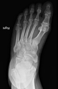
Os vesalianum pedis
Published
23 May 2018
79% complete
X-ray
Case
Accessory hemiazygos vein draining into left brachiocephalic vein

Published
13 Jun 2018
78% complete
CT
Annotated image
Case
Blind-ending branch of a bifid ureter

Published
20 Jun 2018
88% complete
CT
Article
Accessory anterior inferior tibiofibular ligament
The accessory anterior inferior tibiofibular ligament (accessory AITFL), also known as Bassett's ligament, is an anatomical variant present in many ankles. Pathological thickening of the accessory ligament is seen in the setting of inversion injury that causing the pain due to mild anterior inst...
Case
Duplicated middle cerebral artery

Published
20 Aug 2018
66% complete
DSA (angiography)
Case
Os infranaviculare

Published
22 Aug 2018
66% complete
X-ray
Article
Retro-aortic left brachiocephalic vein
The retro-aortic left brachiocephalic vein is a rare vascular variant where the left brachiocephalic vein passes more inferiorly through the superior mediastinum, coursing inferior to the aortic arch and posterior to the ascending aorta to join the right brachiocepahilc vein forming the superior...
Case
Aberrant left main coronary artery (ALMCA) arising from the right sinus with interarterial course

Published
21 Sep 2018
95% complete
CT
Article
Adductor minimus muscle
The adductor minimus muscle is a small, variably present muscle in the medial compartment of the thigh.
Terminology
Due to confusion about this muscle over the years it has ended up with a variety of names, which include pars lateralis, adductor quartus muscle, premier faisceau du grand addu...
Article
Accessory flexor digitorum longus muscle
The accessory flexor digitorum longus muscle is an accessory muscle in the deep posterior compartment of the leg with a reported prevalence of 6-8%. Unilateral muscles are more common although bilateral cases have been reported.
Summary
origin: variable; either the medial margin of the tibia a...
Article
McDonald and McClellan's classification of crossed renal ectopias
McDonald and McClellan classified crossed renal ectopia into four types 1:
bilateral crossed renal ectopia without fusion
unilateral crossed renal ectopia
bilaterally crossed renal ectopia: represents 90% of all crossed ectopias and includes crossed fused renal ectopia
crossed unfused rena...
Case
Cervical thymus

Published
26 Feb 2019
79% complete
Ultrasound
Article
Accessory abductor digiti minimi muscle (hand)
An accessory abductor digiti minimi (ADM) muscle is the most common accessory muscle of the hand and wrist, found in 24% of individuals on the hypothenar eminence. When present it is one of the intrinsic muscles of the hand.
Summary
origin:
antebrachial fascia passing anteriorly to Guyon cana...
Article
Chiari network
A Chiari network refers to a filamentous, weblike structure in the right atrium that results from incomplete resorption of the embryonic sinus venosus. It is an uncommon anatomical variant.
Epidemiology
Prevalence estimates for the general population vary widely, ranging from 2%-10% of randoml...
Case
Castellvi classification - illustrations

Published
25 Sep 2019
16% complete
Diagram
Case
Tracheal bronchus

Published
08 Oct 2019
92% complete
CT
Case
Hangman fracture with incidental arcuate foramen

Published
04 May 2021
85% complete
X-ray
Case
Chiari I malformation with hydrosyringomyelia

Published
21 Nov 2019
93% complete
MRI
Case
Accessory soleus muscle

Published
15 Apr 2020
89% complete
MRI