
Items tagged “variant”
426 results found
Article
Annular pancreas
Annular pancreas is a morphological anomaly that results in pancreatic tissue completely or incompletely encircling the duodenum. This condition can cause duodenal obstruction and is therefore important to recognize, as radiologists are frequently the first to make the diagnosis.
Epidemiology
...
Article
Mesiodens
A mesiodens (plural: mesiodentes) is the most common supernumerary tooth and is located in the palatal midline between the two maxillary central incisors.
Epidemiology
It is rare with an estimated prevalence of ~1% (range 0.09 to 2.2%) 3. There is an increased male predilection with a M:F rati...
Case
Trapezium exostosis

Published
22 Feb 2012
78% complete
MRI
Article
Meckel diverticulum
Meckel diverticulum is a congenital intestinal diverticulum due to fibrous degeneration of the umbilical end of the omphalomesenteric (vitelline) duct that occurs around the distal ileum. It is considered the most common structural congenital anomaly of the gastrointestinal tract.
Epidemiology
...
Article
Circumaortic left renal vein
Circumaortic left renal vein, also known as circumaortic renal collar is an anomaly of left renal vein when a supernumerary or accessory left renal vein passes posterior to the aorta, apart from the normal renal vein passing anterior to the aorta. This anomaly is potentially hazardous, if unreco...
Case
Bilateral persistent median artery of the forearm with unilateral bifid median nerve

Published
12 Apr 2012
74% complete
Ultrasound
Article
Retroaortic left renal vein
Retroaortic left renal vein (RLRV) is a normal anatomical variant where the left renal vein is located between the aorta and the vertebra and drains into the inferior vena cava.
Its recognition is important in order to avoid complications during retroperitoneal surgery or interventional procedu...
Article
Renal vein anomalies
There are several variations in renal venous anatomy. Some of these are specific to the left renal vein.
Left renal vein anomalies are generally classified into four types 2:
type I
the ventral preaortic limb of the left renal vein is obliterated, but the dorsal retroaortic limb persists and...
Case
Abnormal renal vascular impression on intravenous pyelogram

Published
07 May 2012
60% complete
X-ray
Article
Empty sella
An empty sella, also known as an empty pituitary fossa, refers to the appearance of the sella turcica when the pituitary gland appears shrunken or invisible and CSF fills the space instead. It is commonly an incidental finding of no clinical significance, but there exists a well-established asso...
Article
Ectopic posterior pituitary
An ectopic posterior pituitary reflects a disruption of normal embryogenesis of the posterior pituitary and is one of the more common causes of pituitary dwarfism. Although it can be an isolated abnormality, numerous other congenital central nervous system malformations have been identified. Whe...
Case
Persistent trigeminal artery
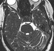
Published
18 May 2012
91% complete
Annotated image
Case
Os supratrochleare dorsale

Published
18 Jun 2012
72% complete
X-ray
Article
Lunotriquetral coalition
A lunotriquetral coalition, also known as lunotriquetral fusion or synostosis, is a type of carpal coalition and represents a congenital lack of separation of the lunate and triquetral bones of the carpus.
Terminology
The term coalition is preferred over fusion for congenital coalitions, as d...
Case
Azygos continuation of the inferior vena cava

Published
11 Jul 2012
65% complete
CT
Case
Left sided superior vena cava

Published
11 Jul 2012
80% complete
CT
Article
Anterior angulation of the coccyx
Anterior angulation of the coccyx may be a normal variant but poses a diagnostic challenge for those considering coccygeal trauma 1.
Classification
Six types of coccyx have been described 2,3:
type I: the coccyx is curved slightly forward, with its apex pointing caudally (~70%)
type II: the ...
Case
Persistent trigeminal artery

Published
20 Jul 2012
77% complete
MRI
Case
Polysplenia syndrome

Published
24 Jul 2012
98% complete
CT
Case
Bicarotid trunk and aberrant right subclavian artery

Published
27 Jul 2012
80% complete
CT
MRI


 Unable to process the form. Check for errors and try again.