


769 results found
Case
Patellar tendon rupture

Published
29 Jan 2024
88% complete
X-ray
Case
Limbus vertebra

Published
13 Nov 2023
74% complete
CT
Case
Buffalo (illustration)

Published
22 May 2023
25% complete
Diagram
Case
Achilles tendon rupture

Published
16 Feb 2023
91% complete
X-ray
Ultrasound
Case
Giant cell tumor with aneurysmal bone cyst-like changes
Published
13 Oct 2022
89% complete
MRI
CT
Case
Brachymetatarsia

Published
08 Jun 2022
79% complete
X-ray
Case
Epidural spinal cord compression scale (illustration)

Published
25 May 2021
25% complete
Diagram
Case
Schmid metaphyseal dysplasia

Published
17 Aug 2020
62% complete
CT
Case
Gouty tenosynovitis

Published
15 Aug 2020
72% complete
Ultrasound
Case
Rheumatoid nodular tendon sheath disease

Published
12 Aug 2020
57% complete
Ultrasound
Case
Gout - wrist

Published
26 Sep 2019
92% complete
CT
MRI
Case
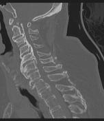
Dens fracture and atlantoaxial subluxation

Published
20 Jun 2019
69% complete
X-ray
Case
Synovial cyst

Published
29 Dec 2018
83% complete
MRI
Case
Facet joint fracture treated with ACDF and follow-up

Published
25 Jun 2018
85% complete
MRI
CT
Fluoroscopy
Case
Hypoxic ischemic injury, diffuse axonal injury and atlanto-occipital dislocation

Published
20 Jun 2018
95% complete
CT
X-ray
MRI
Case
Discitis osteomyelitis

Published
05 Jun 2018
71% complete
MRI
Case
Fibrous dysplasia (polyostotic)

Published
03 Jun 2018
92% complete
MRI
Nuclear medicine
X-ray
Case
Ossification of the posterior longitudinal ligament and DISH
Published
03 Jun 2018
92% complete
CT
Case
Epidural abscess

Published
03 Jun 2018
83% complete
MRI
Case
Sail boat (photo)

Published
29 May 2018
19% complete
Case
Fused hemivertebra

Published
20 May 2018
81% complete
CT
MRI
Case
Blast injury to hand
Published
18 May 2018
79% complete
X-ray
Case
Currarino syndrome

Published
18 Apr 2018
83% complete
MRI
Case
Tuberous sclerosis (multisystem involvement)

Published
29 May 2017
92% complete
CT
X-ray
Ultrasound
MRI
Case
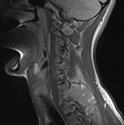
Discitis osteomyelitis - cervical spine

Published
01 Jan 2017
89% complete
MRI
Case
Coronal and sagittal balance

Published
28 Nov 2016
32% complete
Diagram
Case
Cobb angle measurment
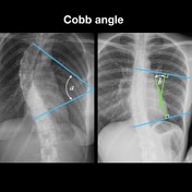
Published
22 Nov 2016
29% complete
Annotated image
Case
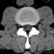
Extruded thoracic disc

Published
08 Nov 2016
84% complete
MRI
CT
Case
Radial neck fracture

Published
04 Nov 2016
85% complete
X-ray
Case
Pelvic fractures, hematoma and pear shaped bladder

Published
20 Oct 2016
92% complete
X-ray
CT
Case
Cisterna chyli

Published
20 Oct 2016
73% complete
Annotated image
MRI
Case
Atlanto-occipital and atlantoaxial distraction

Published
17 Oct 2016
73% complete
MRI
CT
Case
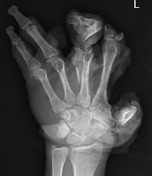
Carpometacarpal dislocation

Published
28 Sep 2016
86% complete
CT
X-ray
Case
Brachial plexus (normal)

Published
28 Sep 2016
58% complete
MRI
Case
Epidural abscess

Published
27 Jul 2016
86% complete
MRI
Case
Vertebral metastases (breast cancer)

Published
23 Jul 2016
92% complete
MRI
CT
Case
Fibrous dysplasia

Published
23 Jun 2016
84% complete
CT
X-ray
Case
Normal cervical spine MRI - including Dixon sequences

Published
02 Mar 2016
53% complete
MRI
CT
Case
Normal wrist CT

Published
18 Feb 2016
65% complete
CT
Case
Normal flexion and extension cervical spine x-rays

Published
27 Jun 2015
63% complete
X-ray
Case
Normal lumbar spine MRI

Published
24 Jun 2015
48% complete
MRI
Case
Elbow dislocation

Published
31 May 2015
66% complete
X-ray
Case
Pyoderma gangrenosum (photo)

Published
19 May 2015
25% complete
Photo
Case
Pelvic tear drop

Published
18 May 2015
22% complete
Annotated image
Case
Acetabular angle

Published
18 May 2015
32% complete
Annotated image
Case
Intervertebral disc disease nomenclature (illustration)
Published
18 May 2015
32% complete
Annotated image
Case
Sagittal localization of disc disease (annotated image)

Published
18 May 2015
32% complete
Annotated image
Case
Disc herniation nomenclature - axial (annotated image)

Published
18 May 2015
32% complete
Annotated image
Case
Vertebral column CT anatomy (annotated image)

Published
18 May 2015
22% complete
Annotated image
Case
Renal osteodystrophy and brown tumors

Published
16 May 2015
69% complete
Annotated image
Case
Popcorn (photo)

Published
16 May 2015
25% complete
Photo
Case
Chondrosarcoma (histology)

Published
16 May 2015
29% complete
Pathology
Case
Zebra (photo)

Published
16 May 2015
25% complete
Photo
Case
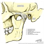
Temporomandibular joint

Published
16 May 2015
24% complete
CT
Case
Inca bone

Published
16 May 2015
21% complete
CT
Case
Normal thigh

Published
16 May 2015
30% complete
CT
Case
Convolutional markings

Published
16 May 2015
24% complete
CT
Case
Orbit (illustration)

Published
16 May 2015
19% complete
Annotated image
Case
Normal acromioclavicular joint

Published
16 May 2015
16% complete
X-ray
Case
Malleus (illustration)

Published
16 May 2015
19% complete
Diagram
Case
Temporalis muscle
Published
16 May 2015
33% complete
MRI
Case
TMJ - retrodiscal area (annotated MRI)

Published
16 May 2015
16% complete
Annotated image
Case
Muscles of mastication (annotated image)

Published
16 May 2015
25% complete
Annotated image
Case
Junction of intermediate zone and posterior band

Published
16 May 2015
30% complete
MRI
Case
Pterygoid muscles - illustration

Published
16 May 2015
29% complete
Diagram
Case
Pterygoid fovea - illustration

Published
16 May 2015
25% complete
Case
Temporalis muscle (illustration)

Published
16 May 2015
19% complete
Diagram
Case
Mandibular fractures (illustration)

Published
16 May 2015
19% complete
Diagram
Case
Condylar process fractures (illustration)

Published
16 May 2015
22% complete
Diagram
Case
Temporomandibular joint (illustration)
Published
16 May 2015
25% complete
Diagram
Case
Wormian bones (photo)

Published
16 May 2015
25% complete
Photo
Case
Bony orbit (photo)

Published
15 May 2015
19% complete
Photo
Case
Facet joint injection - Scotty dog

Published
14 May 2015
19% complete
Fluoroscopy
Case
Celery stalk (photo)

Published
13 May 2015
29% complete
Photo
Case
Pott disease
Published
13 May 2015
22% complete
X-ray
Case
Bilateral distal clavicular erosion

Published
13 May 2015
88% complete
X-ray
Case
Sacroiliitis - grade III

Published
13 May 2015
44% complete
X-ray
Case
Normal sacroiliac joint

Published
13 May 2015
19% complete
X-ray
Case
Distal femoral avascular necrosis

Published
12 May 2015
59% complete
MRI
Case
Doughnut (photo)

Published
12 May 2015
29% complete
Photo
Case
Banana (photo)

Published
12 May 2015
25% complete
Photo
Case
Medusa (photo)

Published
12 May 2015
25% complete
Photo
Case
Palm tree (photo)

Published
12 May 2015
25% complete
Photo
Case
Nail-patella syndrome

Published
12 May 2015
47% complete
Annotated image
Case
Hyperostosis frontoparietalis

Published
12 May 2015
74% complete
CT
Case
Hangman fracture

Published
12 May 2015
54% complete
X-ray
Case
Le Fort fracture - type II

Published
12 May 2015
62% complete
CT
Case
Cervical canal stenosis

Published
11 May 2015
48% complete
MRI
Case
Orbital blow-out fracture - medial

Published
11 May 2015
65% complete
CT
Case
Orbital blow-out fracture

Published
11 May 2015
65% complete
CT
Case
Sacroiliitis - grade IV

Published
10 May 2015
57% complete
X-ray
Case
Acromioclavicular joint injury - type III

Published
10 May 2015
63% complete
X-ray
Case
Persistant ulnar styloid

Published
10 May 2015
38% complete
X-ray
Case
Hyperostosis frontalis interna

Published
10 May 2015
68% complete
CT
ADVERTISEMENT: Supporters see fewer/no ads


 Unable to process the form. Check for errors and try again.