


39 results found
Case
Falx cerebri - Gray's anatomy illustration

Published
24 Mar 2015
45% complete
Diagram
MRI
Case
Brainstem nuclei - dorsal section (Gray's illustration)

Published
17 May 2015
44% complete
Diagram
Case
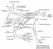
Facial nerve - Gray's anatomy illustration
Published
10 May 2015
41% complete
Diagram
Case
Brainstem - Gray's anatomy illustration

Published
17 May 2015
41% complete
Diagram
Case
Diagram of ventricular system

Published
16 May 2015
38% complete
Diagram
Case
Short association fibers - Gray's anatomy illustration
Published
26 Mar 2015
38% complete
Annotated image
Case
Cerebellum (sagittal) - Gray's anatomy illustration

Published
16 May 2015
38% complete
Diagram
Case
Midbrain (axial) showing tectum and tegmentum

Published
16 May 2015
38% complete
Diagram
Case
Coronal brain through 3rd ventricle

Published
16 May 2015
38% complete
Diagram
Case
Coronal brain in front of pons - Gray's anatomy illustration

Published
16 May 2015
38% complete
Diagram
Case
Cerebral arterial supply to the brain - Gray's anatomy illustration

Published
16 May 2015
38% complete
Diagram
Case
Brainstem sensory nuclei - Gray's anatomy illustration

Published
17 May 2015
35% complete
Diagram
Case
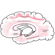
Association fibers (Gray's illustration)

Published
03 Apr 2015
35% complete
Diagram
Case
Brainstem motor nuclei - Gray's anatomy illustration

Published
17 May 2015
35% complete
Diagram
Case
Brainstem - Gray's anatomy illustration

Published
17 May 2015
32% complete
Diagram
Case
Brainstem: dorsal - Gray's anatomy illustration

Published
17 May 2015
32% complete
Diagram
Case
Intracranial ventricles - Gray's anatomy illustration

Published
18 May 2015
32% complete
Diagram
Case
Brainstem: ventral - Gray's anatomy illustration

Published
17 May 2015
32% complete
Diagram
Case
Cisterns and CSF flow - Gray's anatomy illustration

Published
16 May 2015
29% complete
Diagram
Case
Brain (sagittal section) - Gray's anatomy illustration

Published
16 May 2015
29% complete
Diagram
Case
Optic pathways - Gray's anatomy illustration

Published
14 May 2015
29% complete
Diagram
Case
Brainstem nuclei and their connections - Gray's anatomy illustration

Published
16 May 2015
29% complete
Diagram
Case
Epithalamus - Gray's anatomy illustration

Published
16 May 2015
29% complete
Diagram
Case
Meckel cave - Gray's anatomy illustration

Published
15 Mar 2011
29% complete
Diagram
Case
Corpora quadrigemina - Gray's anatomy illustration

Published
08 May 2015
29% complete
Diagram
Case
Midbrain (axial)

Published
16 May 2015
29% complete
Diagram
Case
Midbrain (axial)

Published
16 May 2015
29% complete
Diagram
Case
Basal ganglia location - Gray's anatomy illustration

Published
16 May 2015
29% complete
Diagram
Case
Pituitary region (illustration)

Published
18 May 2015
29% complete
Diagram
Case
Subarachnoid cistern (illustration)

Published
16 May 2015
25% complete
Diagram
Case
Spinal cord meninges (illustration)

Published
16 May 2015
25% complete
Diagram
Case
Base of skull (illustration)

Published
16 May 2015
25% complete
Diagram
Case
Midbrain (level of superior colliculi)

Published
16 May 2015
25% complete
Diagram
Case
Midbrain (level of inferior colliculi)

Published
16 May 2015
25% complete
Diagram
Case
Ventricles superimposed on lateral brain

Published
16 May 2015
25% complete
Diagram
Case
Pituitary gland - Gray's anatomy illustration

Published
16 May 2015
25% complete
Diagram
Case
Pituitary and vascular supply - Gray's anatomy illustration

Published
16 May 2015
25% complete
Diagram
Case
Vagus nerve illustrations - Gray's anatomy illustrations

Published
08 Jul 2013
25% complete
Diagram
Case
Medulla oblongata

Published
31 Dec 2013
22% complete
Diagram
ADVERTISEMENT: Supporters see fewer/no ads


 Unable to process the form. Check for errors and try again.