Phrenic nerve (Gray's illustration)
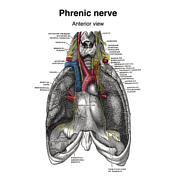
Brachial plexus (Gray's illustrations)

Suboccipital nerves (Gray's illustration)

Posterior sacral nerves (Gray's illustration)

Spinal nerve roots (Gray's illustrations)

Dermatomes (Gray's illustration)

Cutaneous spinal nerves of the upper limb (Gray's illustrations)

Cutaneous spinal nerves of the lower limb (Gray's illustrations)

Bilateral SUFE - different phases

Bilateral fifth metatarsal base fractures

Origins of the extraocular muscles (Gray's illustration)

Internal features of the lateral ventricles (Gray's illustrations)

Innervation of the medial and lateral recti muscles (Gray's illustration)

Hippocampus (Gray's illustration)

Tela choroidea and choroid plexus of lateral ventricles (Gray's illustration)

Internal capsule fibres (Gray's illustration)

Fornix (Gray's illustration)

Corpus striatum (Gray's illustration)

Corona radiata (Gray's illustration)

Basal ganglia (Gray's illustrations)

Normal cardiac CT (volume render)

Lateral talar process avulsion fracture

Pelvic veins (Gray's illustration)

Superficial abdominal wall veins (Gray's illustration)

Enchondroma with pathological fracture and incidental exostosis

Neck veins (Gray's illustration)

Implantable contraceptive device

Dural venous sinuses (Gray's illustrations)

Veins of the scrotum (Gray's illustration)

Female reproductive tract vessels (Gray's illustration)

Superficial veins of the lower limb (Gray's illustration)

Veins of the axilla (Gray's illustration)

Tongue vessels (Gray's illustration)

Vertebral venous plexuses (Gray's illustrations)

Thyroid veins (Gray's illustration)

Superficial veins of the elbow (Gray's illustration)

Cavernous sinus (Gray's illustration)

Superficial veins of the hand (Gray's illustration)

Popliteal vein (Gray's illustration)

Orbital veins (Gray's illustration)

Internal cerebral veins (Gray's illustration)

CT angiogram head sagittal - labelling questions

CT angiogram head coronal - labelling questions

CT foot and ankle coronal - labelling questions

CT foot and ankle axial - labelling questions

CT foot and ankle sagittal - labelling questions

Ischaemic bowel due to internal hernia with perforation
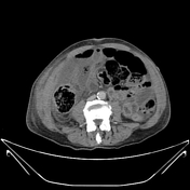
Hereditary multiple exostosis

CT wrist axial - labelling questions

CT wrist sagittal - labelling questions

CT wrist coronal - labelling questions

MRI pituitary gland coronal T1 post contrast - labelling questions

MRI pituitary gland sagittal T1 post contrast - labelling questions

Romanus lesions - ankylosing spondylitis

CT temporal bone sagittal - labelling questions

CT temporal bone coronal - labelling questions

CT angiogram abdomen/pelvis sagittal - labelling questions

Accessory PCA arising from the terminal ICA

Normal CTA abdomen and pelvis (female)

Distal fibular and fifth metatarsal base fractures

CT angiogram abdomen/pelvis coronal - labelling questions

CT angiogram abdomen/pelvis axial - labelling questions

MRI head axial T2 - labelling questions

MRI head sagittal T1 - labelling questions

Anal triangle (illustration)

Urogenital triangle (diagrams)

CT cervical spine axial - labelling questions

Female perineal muscles (Gray's illustration)

Male perineal muscles (Gray's illustration)

Male perineal fascia (Gray's illustration)

Male pelvic and perineal fascia (Gray's illustrations)

Levator ani (Gray's illustration)

CT cervical spine coronal - labelling questions

CT cervical spine sagittal - labelling questions

Normal hip x-ray

Clavicle x-ray - labelling questions

Fractured clavicle and bent ORIF cannulated screw

Teeth anatomy - labelling questions
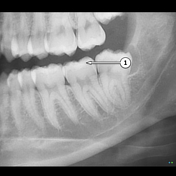
OPG - labelling questions

Truncal venous development (Gray's illustration)

Hepatic venous development (Gray's illustration)

Dural venous development (Gray's illustrations)

Carotid artery development (Gray's illustration)
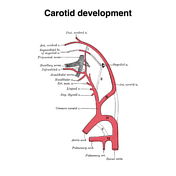
Sinus venosus development (Gray's illustration)

Aortic arches (Gray's illustration)

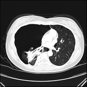
Pneumothorax due to lymphangioleiomyomatosis
Superior ophthalmic vein thrombosis

Cricothyroidotomy tube for severe epiglottitis

Ankle and foot bone infarcts secondary to alcoholism

Femoral canal (Gray's illustration)
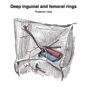
Femoral triangle and sheath (Gray's illustration)

Internal pudendal artery (Gray's illustrations)

Subdural haemorrhage on CT perfusion

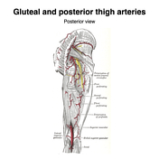
Gluteal arteries (Gray's illustration)
ADVERTISEMENT: Supporters see fewer/no ads


 Unable to process the form. Check for errors and try again.