Odontoid fracture (Anderson and D'Alonzo type 3, Roy-Camille type 1)

Odontoid fracture (Anderson and D'Alonzo type 2, Roy-Camille type 3)

Atlas (type 3b subtype 1) and axis (Anderson and D'Alonzo type 3, Roy-Camille type 2) fractures

Infected ICA pseudoaneurysm
Essex-Lopresti fracture-disclocation

Occipital condyle fracture (type 1) and atlas transverse process fracture (type 5)

Occipital condyle fracture (type 2) with extension into clivus

Bilateral occipital condyle fracture (type 2)

Occipital condyle fracture (type 3)

Occipital condyle fracture (type 1)

Bilateral occipital condyle fractures (type 3)

Wrist CPPD

Atlanto-occipital dissociation (Traynelis type 1), C2 teardrop fracture, C6/7 facet joint dislocation

Atlanto-occipital dissociation - Traynelis type 1

Lester Jones tube
Normal foot CT (with 3D VRTs)

Gehweiler classification of atlas fractures (diagrams)

Roy-Camille classification of C2 odontoid fractures (diagrams)

Anderson and D'Alonzo classification of C2 odontoid fractures (diagrams)

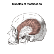
Muscles of mastication (Gray's illustration)
Anderson and Montesano classification of occipital condyle fractures (diagrams)

Traynelis classification of atlanto-occipital dissociation (diagrams)

Talonavicular dislocation

Subtle maxillary sinus fracture

Incidental large faecolith

Fibroid red degeneration in pregnancy

Delayed traumatic splenic pseudoaneurysms

Attempted bifid distal phalanx of the thumb

Windswept knees

Uterine version and flexion (diagrams)

Thoracolumbar injury classification and severity score (table)
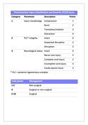
Subaxial cervical spine injury classification (table)

ECG gating (illustration)

Talar neck fracture - Hawkins type 3

Splenic injury - grade 5

Medial malleolar and lateral talar dome fractures

Lymphatics of the thorax and abdomen (Gray's illustration)

Pelvic lymphatics (Gray's illustration)

Lymphatics of the midgut (Gray's illustrations)

Bell clapper (photo)

Bell clapper deformity (diagram)

Salter Harris 4 fracture of index finger middle phalanx

Lymphatics of the stomach and foregut (Gray's illustrations)

Lymphatics of the colon (Gray's illustration)

Lymphatics of the bladder (Gray's illustration)

Lymphatics of the prostate (Gray's illustration)

Lymphatics of the uterus (Gray's illustration)

Lymphatics of the tracheobronchial tree (Gray's illustration)

Lymphatics of the lower limb (Gray's illustration)

Lymphatics of the popliteal fossa (Gray's illustration)

Female mammary gland (Gray's illustration)

Lymphatics of the breast and axilla (Gray's illustration)

Extensive rib plating for flail segment

Ankle and foot interosseous ligaments (Gray's illustrations)

Lymphatics of the upper limb (Gray's illustration)

Lymphatics of the tongue (Gray's illustration)

Lymphatics of the face (Gray's illustration)

Lymphatics of the pharynx (Gray's illustration)

Lymphatics of head and neck (Gray's illustration)

Patellar tendon rupture

Thoracic and right lymphatic ducts (Gray's illustration)

Menstrual cup on trauma imaging

Hepatic abscess

Plantar ligaments of the foot (Gray's illustration)

Lateral talocrural ligaments (Gray's illustration)

Ankle and foot ligaments (Gray's illustration)

Subtalar ligaments (Gray's illustration)

Patella (Gray's illustration)

Knee joint capsule (Gray's illustration)

Hoffa's fat pad (Gray's illustration)

Homolateral Lisfranc injury

Knee menisci (Gray's illustration)
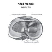
Internal knee ligaments (Gray's illustrations)

Knee ligaments (Gray's illustration)

Normal pelvis CT angiogram

Swallowed pen

Hip joint capsule (Gray's illustration)

Ligamentum teres of the hip (Gray's illustrations)
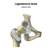
Hip capsular ligaments (Gray's illustration)

Ligaments of the fingers (Gray's illustration)

Triangular fibrocartilage complex (Gray's illustration)

Wrist ligaments (Gray's illustrations)

Proximal radio-ulnar joint ligaments (Gray's illustration)

Shoulder bursa (Gray's illustration)

Sternoclavicular joint (Gray's illustration)

Inguinal and lacunar ligaments (Gray's illustration)

Pelvic ligaments (Gray's illustrations)

CT angiogram head axial - labelling questions

Normal tongue base MRI

Tectorial membrane and C0-1-2 ligaments (Gray's illustration)

Atlanto-odontoid joint (Gray's illustration)

Posterior atlanto-occipital membrane (Gray's illustration)

Anterior atlanto-occipital membrane (Gray's illustration)

Ligamentum flavum (Gray's illustration)

ADVERTISEMENT: Supporters see fewer/no ads


 Unable to process the form. Check for errors and try again.